Fig. 1
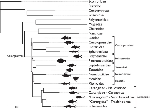
Hypotheses of relationships among the Carangiformes based on the following molecular studies: Smith and Wheeler, 2006; Li et al., 2011; Near et al., 2013; Betancur-R. and Ortí, 2014; Harrington et al., 2016; Mirande, 2016; Smith et al., 2016; Rabosky et al., 2018. Latidae, Polynemidae, and members of the infraorder Pleuronectoideo are denoted by a triangle, a circle, and one or more stars, respectively, to highlight the conflicting hypotheses of carangiform relationships across previous DNA-based studies.
Fig. 2
Hypotheses of relationships from partitioned likelihood analysis of carangiform fishes and outgroup taxa. The dataset included 201 discrete morphological characters and 463 ultraconserved element loci. Cladogram condensed to show the relationships among families and larger groups sampled. See Data Accessibility for tree file.
Fig. 3
Cladogram from partitioned likelihood analysis of carangiform fishes. Dataset included 201 discrete morphological characters and 463 ultraconserved element loci. Cladogram restricted to only the Carangiformes and Synbranchiformes. Morphological characters optimizing onto each node are represented by a circle with the corresponding character number listed above and corresponding character state below. Circles with black fill-in are unique and unreversed characters. Circles with white fill-in are inferred to have evolved multiple times in the cladogram.
Fig. 4

Morphological variation in support of the relationships among the Centropomoidei. Images of cleared and stained specimens fluorescing under royal blue light. Presence of rostral extension of external process on maxilla (character 361)—(A) Centropomus undecimalis (FMNH 77806), arrow, lateral view of right maxilla, scale bar = 5 mm; (B) Centropomus undecimalis (FMNH 77806), arrow, dorsal view of right maxilla, scale bar = 5 mm; (C) Sphyraena barracuda (FMNH 74209), arrow, lateral view of right maxilla, scale bar = 5 mm; (D) Sphyraena barracuda (FMNH 74209), arrow, dorsal view of right maxilla, scale bar = 5 mm. Enlargement of the second abdominal neural spine (character 1621)—(E) Lates calcarifer (AMNH 37839), arrow, right lateral view, scale bar = 1 mm; (F) Centropomus undecimalis (FMNH 77806), arrow, right lateral view, scale bar = 2.5 mm; (G) Lactarius lactarius (KUI 41405), arrow, right lateral view, scale bar = 5 mm. Possession of elongated, fang-like teeth in the oral jaws (character 261) and oral teeth that are ankylosed to the premaxilla and dentary (character 271)—(H) Sphyraena idiastes (SIO 15–182), lateral view of right premaxilla, scale bar = 1 mm; (I) Lactarius lactarius (KUI 41405), lateral view of right premaxilla, scale bar = 1 mm; (J) Lactarius lactarius (KUI 41405), lateral view of right dentary, scale bar = 1 mm.
Fig. 5
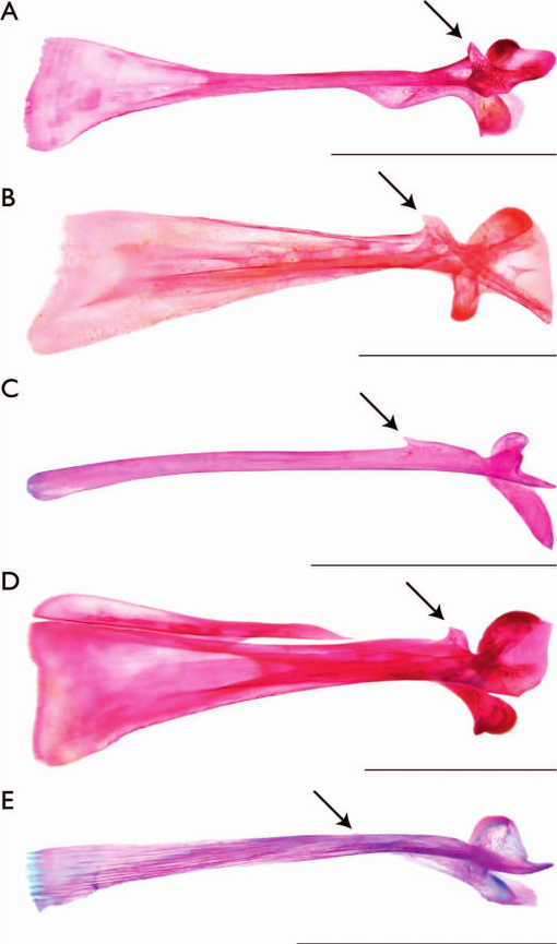
Morphological variation in support of the relationships among carangiform taxa not included in the Centropomoidei. Images of cleared and stained specimens under white or daylight LED light. Dorsally directed external process on maxilla (character 341)—(A) Polydactylus sexfilis (CAS 50911), arrow, lateral view of right maxilla, scale bar = 5 mm; (B) Scophthalmus aquosus (KUI 30388), arrow, lateral view of right maxilla, scale bar = 5 mm; (C) Toxotes jaculatrix (KUI 42174), arrow, lateral view of right maxilla, scale bar = 5 mm; (D) Caranx hippos (KUI 30383), arrow, lateral view of right maxilla, scale bar = 5 mm; (E) Coryphaena hippurus (FMNH 48561), arrow, lateral view of right maxilla, scale bar = 5 mm.
Fig. 6
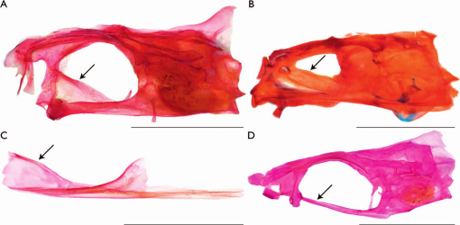
Morphological variation in support of the relationships among the Polynemoidei. Images of cleared and stained specimens under white or daylight LED light. Anterodorsal expansion of the parasphenoid (character 81)—(A) Filimanus similis (LACM 38134.017), arrow, left lateral view of neurocranium, scale bar = 1 cm; (B) Psettodes erumei (ANSP 214777), arrow, left lateral view of neurocranium, scale bar = 5 mm; (C) Polydactylus sexfilis (CAS 50911), arrow, left lateral view of isolated parasphenoid, scale bar = 5 mm. Absence of anterodorsal expansion of the parasphenoid (character 80)—(D) Toxotes jaculatrix (FMNH 69510), arrow, left lateral view of neurocranium, scale bar = 1 cm.
Fig. 7
Morphological variation in support of the relationship among the Carangoidei, Menoidei, Nematistioidei, and Toxotoidei. Images of cleared and stained specimens under white or daylight LED light. Dentition present on the basihyal (character 641)—(A) Toxotes blythii (KUI 42173), dorsal view of basihyal, scale bar = 1 mm; (B) Leptobrama muelleri (KUI 41406), dorsal view of basihyal, scale bar = 1 mm; (C) Xiphias gladius (MCZ 55512, Museum of Comparative Zoology, Harvard University, ©2019 President and Fellows of Harvard College), dorsal view of basihyal, scale bar = 1 mm; (D) Oligoplites saurus (KUI 17205), dorsal view of basihyal, scale bar = 1 mm; (E) Coryphaena hippurus (FMNH 48561), dorsal view of basihyal, scale bar = 1 mm; (F) Trachurus trachurus (KUI 19964), dorsal view of basihyal, scale bar = 1 mm.
Fig. 8
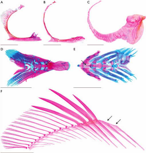
Morphological variation in support of the relationship among the Menoidei, Nematistioidei, and Toxotoidei. Images of cleared and stained specimens under white or daylight LED light. Increased number of circumorbital elements (character 161)—(A) Leptobrama muelleri (KUI 41406), right lateral view, scale bar = 5 mm; (B) Nematistius pectoralis (SIO 12-3085), right lateral view, scale bar = 5 mm; (C) Mene maculata (FMNH 119713), right lateral view, scale bar = 5 mm. Basibranchial two is the anteriormost visible basibranchial when viewed dorsally, as basibranchial one is covered dorsally by the basihyal (character 631)—(D) Toxotes blythii (KUI 42173), arrow, dorsal view of branchial elements, scale bar = 5 mm. Basibranchial one is the anteriormost visible basibranchial with the basihyal anterior to and in series with basibranchials (character 630)—(E) Achirus lineatus (FMNH 113137), arrow, dorsal view of branchial elements, scale bar = 5 mm. Dorsal pterygiophore without associated dorsal spine (character 1351)—(F) Toxotes jaculatrix (FMNH 69510), arrows, right lateral view of dorsal-fin elements, scale bar = 5 mm.
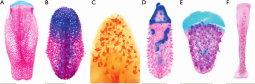
Fig. 9
Morphological variation in support of the relationship among the Menoidei. Lateral flaring of the parasphenoid (character 111)—(A) Mene maculata (KUI 42175), skeletal specimen imaged under white or daylight LED light, ventral view, scale bar = 5 mm; (B) Xiphias gladius (MCZ 55512, Museum of Comparative Zoology, Harvard University, ©2019 President and Fellows of Harvard College), cleared and stained specimen imaged under white or daylight LED light, ventral view, scale bar = 1 mm.
Fig. 10
Representative convergent morphological characters coded in this study. Images of cleared and stained specimens under white or daylight LED light. Presence of hypurostegy (character 1901)—(A) Istiophorus platypterus (SIO 73-269), arrow, right lateral view, scale bar = 1 mm; (B) Mene maculata (FMNH 119713), arrow; right lateral view, scale bar = 5 mm; (C) Scomber japonicus (KUI uncat.), arrow, right lateral view, scale bar = 5 mm. Presence of interdigitation between metapterygoid and quadrate (character 461)—(D) Polydactylus virginicus (FMNH 104648), arrow, medial view of right suspensorium, scale bar = 2.5 mm; (E) Leptobrama muelleri (KUI 41406), arrow, medial view of right suspensorium, scale bar = 2.5 mm.


