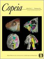
The endocranial space, nerve pattern, and inner ear of a specimen of Uropeltis woodmasoni are described from digital endocasts rendered from high-resolution X-ray computed tomography scans. The cast of the endocranial space reveals short, stout olfactory bulbs as well as general shortening of the brain along the longitudinal axis. The pattern of innervation agrees with descriptions reported for other uropeltid snakes, although this individual exhibits skull asymmetry in the pathway of the glossopharyngeal nerve and the bifurcation of the jugular canal. There are no internal connections between the perilymphatic canal leading to the foramen pseudorotundum, the recessus scala tympani, and the jugular canal, a situation that differs from those of other alethinophidian snakes. The inner ear is compactly packaged with smoothly curved semicircular canals located in close proximity to the vestibule. Semicircular canal size is comparable to those of larger snakes, and the small size of the skull (9.4 mm) coupled with relative enlargement of the sensory regions of the brain and otic capsule is consistent with the morphological pattern of miniaturized vertebrates, although compounding factors, including phylogeny, ontogeny, and variation, are currently not well understood.
How to translate text using browser tools
26 February 2010
Digital Endocasts of the Cranial Cavity and Osseous Labyrinth of the Burrowing Snake Uropeltis woodmasoni (Alethinophidia: Uropeltidae)
Jennifer C. Olori
ACCESS THE FULL ARTICLE