The orchid mantis, Hymenopus coronatus (Insecta: Mantodea), is renown for its visual resemblance to a flower blossom. It has been hypothesised that the ‘flowerlike’ orchid mantis is an aggressive mimic that attracts pollinators as prey items. This is the first study into the morphology of the orchid mantis that explores this widely discussed hypothesis. We quantified color and shape patterns of orchid mantises that are likely to present visual cues to pollinators. We used spectrometry to measure their overall coloration and geometric morphometric techniques to quantify the shape of their ‘petal-like’ mid- and hind-legs. This was done for both juvenile and adult female orchid mantises. To investigate how this stimulus may be perceived by a pollinating insect we investigated within-individual color variation using physiological models of hymenopteran vision. Mantises were found to reflect primarily UV- absorbing white. Visual models indicated that within individuals, different body parts did not contrast highly in color. Femoral lobes showed patterns of bilateral symmetry with juveniles expressing similar patterns of shape variation to adults. The results are used to provide specific and testable hypotheses as to how the morphology of the orchid mantis may constitute a signal directed towards pollinating insects.
Introduction
Animal colors have intrigued scientists for centuries. Some of the earliest applications of evolutionary theory were used to establish the basis that these colors carried functions other than for the novelty of mankind (Wallace 1877). Since then numerous adaptive functions for animal colors have been described including camouflage, sexual signalling, thermoregulation and aposematism (Poulton 1890; Cott 1940).
One of the most commonly discussed functions of color is mimicry. Mimicry theory states that an organism may gain fitness benefits by resembling another unrelated organism (Pasteur 1982). Typically, discussions of mimicry focus on organisms that avoid predation by resembling a distantly related and unpalatable organism. However, there are various other phenomena that benefit from mimetic resemblances (see Pasteur 1982; Ruxton et al. 2004). One of the best known is floral mimicry — the ability of some plants to deceive pollinators into visiting non-rewarding flowers by resembling a rewarding stimulus (Roy & Widmer 1999).
There are various forms of deceptive pollination in flowers (Dafni 1984). In food deceptive floral mimicry, pollinators mistake a nonrewarding flower for a rewarding flower based on visual similarity. The possibility that this same form of deception could occur in animals has been hypothesised yet never tested explicitly. A small number of animals have been suggested as potential flower mimics such as the praying mantis Idolum diabolicum (Varley 1939) and the flatid bugs Flata nigrocincta (Hinde 1902). In most cases little or no data are available and floral mimicry does not appear to uphold as an entirely convincing hypothesis for these animals' morphology. In exception to this, floral mimicry in the orchid mantis, Hymenopus coronatus (Mantodea: Hymenopodidae), remains a plausible and compelling hypothesis.
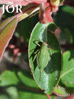
The orchid mantis is native to Indonesia and South East Asia. Little is known of its biology yet it is a well-known, charismatic insect due to its unique morphology. This predatory insect species is characterized by having large, flat expansions of exoskeleton (femoral lobes) on the femur of the mid and hind legs (Fig. 1). They are predominantly white in color and can often have pink or yellow hues. To humans, the juveniles resemble a flower blossom as a result of their four ‘petal-like’ femoral lobes, broad abdomen and bright coloration. This resemblance to a flower blossom has led to the suggestion that juvenile H. coronatus mimic flowers to attract pollinators as prey.
Alfred Russell Wallace first brought the predatory strategy of the orchid mantis to the attention of biologists in 1877 when he recounted a story told by British politician Sir Charles Dilk who, when travelling through Indonesia, was shown a praying mantis that resembled a pink orchid flower (Dilk 1868; Wallace 1889).This incredible tale was soon recounted by others and became featured in a number of classic texts on animal coloration (Wallace 1877; Wood-Mason 1878; Wallace 1889; Poulton 1890). During the Skeat Expedition to the Malaysian Peninsula in 1899, Lord Nelson Annandale observed an orchid mantis perched upon a flower of Melastoma polyanthum (Annandale 1900). His observations of its behaviour overthe following days were published in the Proceedings of the Zoological Society of London. Much of what he observed has been repeated elsewhere (e.g., Shelford 1902; Cott 1940; Stephenson 1946; Edmunds & Brunner 1999 ) and it seems that this publication, based on the observation of a single mantis, is the source of many widely held beliefs about orchid mantis biology.
The orchid mantis commonly features in popular and general natural history texts (e.g., Yong 1976; Edmunds & Brunner 1999; Dawkins 2009) and the hypothesis that the orchid mantis mimics a flower has persisted in the scientific and non-scientific literature despite there being no evidence to support this (e.g., Edmunds & Brunner 1999; Dawkins 2009). Furthermore, there has been no investigation into the morphology of H. coronatus that could give more precise information on how it could constitute a signal to other organisms.

When observing flowers, pollinators respond to contrast between the flower and its background, and patterns within the flower itself. Contrasting color patches, pattern elements and shape outlines can affect the preferences of, and the approach and alighting behaviour ofpollinators (e.g., Lunau 1992; Lehrer et al. 1995; Dafni 1996; Dyer & Chittka 2004; Lunau et al. 2006). If orchid mantises represent a flower-like stimulus then their morphology may consist of a number of color and shape components that combine to form a complex stimulus. The white/pink body coloration and the femoral lobes appear to be derived characteristics of the orchid mantis that contribute to their resemblance to flowers. By quantifying variation in these features we have two main aims; 1 ) to quantify and describe the color and shape of the orchid mantis and 2) to suggest testable hypotheses as to how orchid mantis shape and color components may combine to present a signal directed towards pollinators. Variation in the shape of their femoral lobes is quantified using geometric morphometries. Entire body coloration across visible wavelengths is quantified using spectrophotometry. To investigate how pollinators perceive these colors we employed two physiological models of trichromatic hymenopteran vision. This was done using juvenile female orchid mantises and also the less ‘flower-like’ adult female orchid mantises. By investigating how the body of the orchid mantis is perceived by pollinators at a sensory level we aim to provide an impetus for future research into this phenomenon.
Methods
Orchid mantises are rare animals and large sample sizes are difficult to obtain. Female Hymenopus coronatus used in this study were from captive populations maintained by private insect keepers in Peninsular Malaysia and Singapore. Male orchid mantises in particular are difficult to come by and at the time of this study only female mantises were available in reasonable sample sizes.
We assumed that the dorsal surface of H. coronatus is most likely to be viewed by other organisms. We identified a number of areas on the dorsal surface for which color was measured separately. In adults (n=9) these were the femoral lobes of hind and mid legs, dorsal prothorax and wings. In juveniles (n=15) these were the femoral lobes of hind and mid legs, dorsal prothorax, wing buds and dorsal abdomen surfaces. As juveniles often rest with their abdomen raised or held over their thorax (pers. obs.) we also measured the color of the juveniles' ventral abdomen surfaces, as these may also be visible to other organisms. Color and shape of juvenile and adult mantises were analysed separately.
Spectral reflectance measurements across the visual light spectrum (300–700 nm) were taken from each body part of interest using a spectrometer (Jaz EL-200 with PX2 light source - Ocean Optics Inc. Florida). Reflectance curves were obtained using the average reflectance from 3–5 randomly positioned points on each body part.
If the photoreceptor sensitivities of a hypothetical signal receiver are known, one can calculate the response of these photoreceptors to a reflectance spectrum of interest, when viewed against a given background and under a given illumination spectrum. As varying reflectance spectra will differ in how they excite a system of photoreceptors, a numeric value of chromatic contrast between two colors can be calculated. These contrasts can then be compared to threshold values to infer whether two colors differ enough to be perceived as so by the viewing organism. Where chromatic contrast values are higher than threshold values we predict that the receiver should be able to distinguish between these colors based on their chromaticity. These threshold values can be inferred from behavioural experiments, as in the color hexagon model (Chittka 1992), or from the physiological limitations of photoreceptor cells, as in the receptor noise limited model (Vorobyev & Osorio 1998).
Fig. 2.
Orchid mantis femoral lobe showing placement of landmarks for geometric morphometric analysis (green = fixed, white = semi-sliding).

Fig. 3.
Average percentage reflectance curves ±SD of a) juvenile dorsal abdomens, b) juvenile ventral abdomens, c) juvenile femoral lobe (mid-right), d) juvenile wing buds, e) juvenile prothorax, f) adult femoral lobe (mid-right), g) adult wings and h) adult prothorax.

Fig. 4.
Juvenile female H. coronatus brightness when photographed under normal light (left) when photographed using a UV filter (right).

To test for color contrast within individual orchid mantises from the perspective of pollinators (i.e., chromaticity), we employed two visual models; the receptor noise limited model (Vorobyev & Osorio 1998) and the color hexagon (Chittka 1992). Using these models (see details below) we calculated the chromatic contrast between each body part - ΔS for the receptor noise limited model and chromatic contrast (CC) for the color hexagon - between each of the body parts of individuals.
Calculations for the two models used in this study can be found elsewhere (Kelber et al. 2003 and references therein). We used the photoreceptor sensitivities of honeybees (Menzel & Backhaus 1991) as a hypothetical receiver. As photoreceptor sensitivities vary little in the Hymenoptera (Chittka et al. 1992; Peitsch et al. 1992) this analysis may reflect the sensory biases of hymenopteran pollinators in general. Illumination standard D65 was used as an ambient light spectrum. A background reflectance spectrum was calculated as an average spectrum from a random sample of green leaves found at the University of Malaya Ulu Gombak Field Studies Centre.
Chromatic contrast between body parts was compared to discrimination threshold values (ΔS of 1 for the receptor noise model and CC of 0.05 for the color hexagon). One sample t-tests were used to assess whether average contrast values significantly differed from threshold values. We used the program AVICOL (Gomez 2006) to calculate chromatic contrast values, subsequent analyses were conducted using R 2.14.1 (R Development Core Team 2011).
Geometric morphometries.— The femoral lobes of the mid and hind legs are an important feature of the orchid mantis' flower-like appearance. The four femoral lobes in combination with the broad abdomen in juveniles give the appearance of five petals radiating out from a central point. To investigate any spatial patterning present in the signal generated by mid and hind legs we used geometric morphometrics to investigate shape variation in the femoral lobes, and tested for differences in shape between the four femoral lobes of an individual.
Traditional morphometric techniques characterize morphology in terms of absolute parameters, such as linear distances and areas, or the ratios and angles formed by absolute values. Geometric morphometrics differs in that it summarizes overall shape by quantifying the relative spatial relationships of a number of landmarks (for review see Slice 2007). Shape variables then quantify those variations in morphology that can be attributed to variation in the relative position of identifiable landmarks on a given specimen and are independent of changes in size, position and orientation.
Generalized procrustes analysis is performed on the position of landmarks of a number of specimens to align specimens and generate an average configuration of landmarks. Specimens are scaled to the same limit centroid size and brought into a common arbitrary coordinate system using the average configuration as a reference point. Thus landmark coordinates of specimens superimposed upon the average configuration constitute shape variables rather than raw co-ordinates.
From this coordinate system principal components that describe the variability in positions of corresponding landmarks among specimens can be derived (Adams et al. 2004). For this study we used relative warp analysis to generate relative warp scores, essentially principal components of shape to identify and quantify variation in shape among homologous structures. Patterns of variation in these scores can then be analyzed using traditional statistical methods.
Digital photographs were taken of each of the four femoral lobes of 16 juvenile orchid mantises. The outline of each femoral lobe was digitized using 22 evenly spaced landmarks (3 fixed, 19 sliding semi-landmarks; see Fig. 2) Digitizations were conducted using the program tpsDig 2.16 (Rohlf 2010a). Relative warp (RW) scores were calculated from these digitized specimens using tpsRelw 1.49 (Rohlf 2010b). This process was repeated separately for adult orchid mantis femoral lobes (n=17).
Table 1.
Average AS values of chromatic contrast between juvenile orchid mantis' body parts (FL=femoral lobe) as calculated by the receptor noise limited model. Two-tailed significance values indicated as **<0.05, *<0.001 when testing for significant difference to threshold value of ΔS=1 (One-sample t-test).

Table 2.
Average chromatic contrast (CC) between juvenile female orchid mantis' body parts (FL=femoral lobe) as calculated by the color hexagon model. Two-tailed significance values indicated as **<0.05, *<0.001 when testing for significant difference to threshold value of CC=O.05 (One-sample t-test).

Subsequent analyses of RW scores were performed using R2.14.1 (R Development Core Team 2011 ). RW scores that explained at least 5% of shape variation were then tested for differences between leg types. Paired samples ANOVA was to test whether values for each RW score differed between the femoral lobes of each individual (i.e., mid right — mid left — hind right — hind left), accounting for variation between individuals.
A test was conducted to assess the repeatability of using RW scores for quantifying variation in femoral lobe morphology. RW scores were calculated for repeated digitizations of a sub sample of 10 juvenile legs. Variation in RW scores between specimens was significantly higher than within specimens (F9,20=3.8-245.9, p=<0.01) and repeatability for the first five warp scores was high (0.99 – 0.78).
Results
Color contrast within individuals.— The legs and abdomen of juvenile H. coronatus (n=15), which appear white to humans, were UV absorbing (Fig. 3). The wing buds and prothorax reflected less intensely and the wing buds appeared to have slightly higher reflectance in the UV than other body parts (see Fig. 3d). Fig. 4 shows the orchid mantis photographed using an ultraviolet filter demonstrating that slight reflectance in the UV is restricted to the wing buds.
Both hymenopteran visual models yielded similar results (Tables 1 and 2). Chromaticity varied between body parts yet in most cases any color contrast within individuals was significantly below discrimination threshold values. Contrast between the UV reflecting wing buds and other body parts yielded some of the highest chromatic contrast values, in some cases significantly higher than discrimination threshold values.
Adult H. coronatus (N=6) also reflected light brightly across the human visual spectrum (Fig. 3). There was no evidence of UV reflectance on any body part as was found for the juveniles. Similarly to juveniles the prothorax appeared to reflect less intensely to other body parts. Chromatic contrast within individual adults was mostly either significantly below or not significantly different from discrimination threshold values (Tables 3 and 4). Both the color hexagon model and the receptor noise limited model ranked within individual chromatic contrasts similarly. The wings were overall the most highly chromatically contrasting body part when compared to the legs and prothorax.
Table 3.
Average AS values of chromatic contrast between adult female orchid mantis' body parts (FL= femoral lobe) as calculated by the receptor noise limited model. Two-tailed significance values indicated as **<0.05, *<0.001 when testing for significant difference to threshold value of ΔS=1 (One-sample t-test).

Table 4.
Average chromatic contrast (CC) between adult female orchid mantis' body parts (FL=femoral lobe) as calculated by the color hexagon model. Two-tailed significance values indicated as **<0.05, *<0.001 when testing for significant difference to threshold value of CC=0.05 (One-sample t-test).

Table 5.
Percentage of shape variation in juvenile femoral lobes explained by relative warp scores. Paired samples ANOVAs were used to test whether relative warp score values differed between the four femoral lobes of individuals.

Table 6.
Percentage of shape variation in adult femoral lobes explained by relative warp scores. Paired samples ANOVAs were used to test whether relative warp score values differed between the four femoral lobes of individuals.

Femoral lobe morphology. —The first four RW scores generated by geometric morphometric analysis of juvenile femoral lobes accounted for over 90% of the variation in shape (Table 5). The shape characteristics that RW scores can quantify are often difficult to describe. Thin plate spline deformation grids are often used to visually depict the transformation in shape that is expressed by variation in any given RW score. These diagrams represent variation in RW scores as a deformation of the average landmark coordinates. Figure 5 depicts the main shape characteristics in which juvenile femoral lobes may vary. Paired samples ANOVA found that there were significant within individual differences in femoral lobe shape for RW scores one and three (Table 5 and Fig. 6). Post-hoc testing showed that the mid legs differed significantly in shape to the hind legs for RW score one (Tukey's post-hoc: RW1, z=3.878-4.522, p<0.001). There was no significant difference in RW score one values between left and right side femoral lobes (z=-0.367- -0.108, p>0.5). For RW score three significant differences were only detectable between mid right and hind left femoral lobes (z=3.488, p=0.003), and mid right and hind right femoral lobes (z=3.264, p=0.006).
Over 88% of the variation in adult femoral lobe shape was explained by the first four RW scores (Table 6). Inspection of thin plate spline deformation grids suggests that these RW scores describe shape variables that are very similar to those identified in the juveniles (Fig. 7). Paired samples ANOVAs found significant differences between femoral lobes for RW scores one and three (Table 6 and Fig. 8). As in juveniles, RW score one differed significantly between hind and mid femoral lobes (z=-4.657- -4.103, p<0.0003), but not left and right side (z=-0.0220.523, p>0.9). Significant differences in RW score three were only detected between mid right and hind right legs (z=2.705, p=0.034).
Discussion
Hymenopus coronatus present a primarily UV-absorbing white stimulus to potential signal receivers. Whilst there was some variation in color within individuals, chromatic contrast values were generally quite low. Discrimination threshold values do not represent definite predictions of animal behaviour as the ability of animals to distinguish colors can be context dependant and influenced by factors such as behavioural conditioning and ambient lighting conditions (e.g., Dyer & Chittka 2004; Ings & Chittka 2008).
The shape of femoral lobes differed within individuals. Geometric morphometric analyses uncovered slight differences between the mid and hind legs primarily attributable to the shape variable expressed by RW score one. From examination of the shape characteristics described by RW score one (Fig. 5 ) it appears that hind-leg femoral lobes may have a more elongated form whereas mid-leg femoral lobes are more rounded in shape. This was found in both adult and juvenile mantises. There is, however, a great deal of variation and overlap in these scores (Figs 6, 8). Inspection of thin plate splines suggests that even extreme variations in these RW scores do not produce great amounts of perceivable deformation from a typical femoral lobe shape. It is unlikely that these slight shape differences affect the overall signal emitted by orchid mantises and may reflect developmental differences between mid and hind legs. There was no difference in femoral lobe morphology between right and left legs. With the absence of any color contrast between right and left sides the orchid mantis' body would present a bilaterally symmetrical signal to receivers.
Fig. 5.
Thin plate spline deformation grids visualising shape variation in juvenile femoral lobes as characterized by the first three relative warp scores. Minimum and maximum values of each relative warp score represent the highest values exhibited by individuals in this study.

There are other aspects of H. coronatus coloration that were not investigated in this study. Hymenopus coronatus are known to change between UV-absorbing white and pink color variations throughout their juvenile stages. This pink coloration appears to develop for a few days before and after ecdysis (J.C. O'Hanlon, pers. obs.). Mantises available for this study were primarily white in hue. As such we still know little about the characteristics of mantises with apparently pink hues.
Fig. 6.
Relative warp scores of generated from geometric morphometric analysis of all four femoral lobes of individual juvenile H. coronatus. Symbols indicate femoral lobes from different appendages; mid left leg (•), mid right leg (▴), hind left leg ( ) and hind right leg (▵).

In juveniles a thin green band across the prothorax, and five red/brown anterolateral stripes on the dorsal surface of the abdomen are consistent and conspicuous features of their coloration. In his observations of a wild orchid mantis, Annandale (1900) elaborated on possible functions of these features. He suggested the green band on the prothorax served as a disruptive band that could disrupt the perception of the head of the mantis. He also suggested that the stripes on the abdomen might give the appearance of a withering petal, or specifically resemble the stripes of an orchid's labellum. Parallel or radiating stripes are common features of signals directed towards pollinators. They feature commonly in flowers and have been noted to also occur in structures such as the entrances to pitcher plants and bee nests (Biesmeijer et al. 2005). Biesmeijer et al. (2005) have suggested that bees innate preferences to radiating stripe patterns have led to the convergent evolution of similar patterns on a range of different structures used as visual cues by pollinators. Whether these other color components have functional effects on orchid mantis foraging and signalling requires further investigation.
Fig. 7.
Thin plate spline visualisations of variation in adult femoral lobe shape as characterized by the three first relative warp scores.

The floral mimicry hypothesis is generally discussed in reference to juvenile orchid mantises. The illusion of floral resemblance appears to be lost in adult orchid mantises due to their long wings extending over the thorax and abdomen. Given that adults still retain femoral lobes and similar UV absorbing white coloration to the juveniles it is possible that they also present an attractive stimulus to pollinators, or the lobes may simply be developmentally fixed such that they are retained into adulthood because they pose no disadvantage. Male orchid mantises are much smaller than females and are also fully winged as adults. Males were not available for this study so comparative assessments of their coloration and morphology could not be made however males also have femoral lobes and are white/pink in color. Floral mimicry or sensory exploitation?—Predators can misclassify animals as inedible plant parts due to the specific resemblance of the prey item to an inedible object (Skelhorn et al. 2010b). The protection the prey item gains from this camouflage strategy, termed ‘masquerade’, is not achieved by blending into their background to avoid detection, it is a strategy which exploits the cognitive processes of experienced predators so that even if the prey item is detected it is erroneously identified as something inedible or uninteresting (Skelhorn et al. 2010a). Although the resemblance of juvenile H. coronatus to flowers has so far been based on human assessments, the apparent sophistication of the resemblance could suggest that a similar process to masquerade may occur in this system. Apollinator having experienced rewarding flowers may then perceive the body of an orchid mantis as a whole, intact flower corolla.
Fig. 8.
Relative warp scores of showing variation in shape between femoral lobes of individual adult H. coronatus. Symbols indicate femoral lobes from different appendages; mid left leg (•), mid right leg (▴), hind left leg () and hind right leg (▵).

It is entirely possible that the fitness benefits of imitating a flower can be attained without a specific resemblance to a model object as in mimicry or masquerade. In a number of orb-web spider species bright color patches on the spider's body (Hoese et al. 2006; Tso et al. 2006; Fan et al. 2009), as well as the presence of web decorations have been shown to attract prey towards the spiders web (for reviews see Herberstein et al. 2000; Walter & Elgar 2012). It has been suggested that the color of body patches and the orientation of web decoration structures emit signals similar to those often associated with food resources in pollinating insects such as bright colors and radiating patterns (Tso et al. 2006; Fan et al. 2009; Cheng et al. 2010). In this case an apparent resemblance to flowers may be more accurately described as a form of sensory exploitation rather than a form of mimicry.
If the orchid mantis' morphology functions as a form of mimicry this implies that the orchid mantis is recognised and cognitively misclassified as a flower. Thus the shape and color features of the orchid mantis' body should combine to emit a complex signal that converges upon a model species. Some have suggested that the resemblance to an orchid flower may be quite specific. Wallace (1889) suggested that the appearance of an orchid with four petals and a broad labellum could be mimicked by the mantis' four legs and broad abdomen, whereas the head and thorax of the mantis resembled the column of an orchid flower. Classical mimicry theory suggests that for pollinators should have previous experience with a rewarding model species thus leading them to misclassify the mimic as a rewarding flower (Dafni 1984). Whether the orchid mantis resembles a particular type of flower is unknown. If they do mimic a particular model species then we would predict that orchid mantis shape and coloration should resemble this species more than others.
It is likely that mimicry and sensory exploitation are not mutually exclusive mechanisms. Our ability to empirically distinguish between the two is limited due our current knowledge of the cognitive processes of organisms in their natural environments. This is particularly relevant in the case of pollinator deception. The dynamics of floral mimicry systems are often highly variable (Roy & Widmer 1999) and the perceptual biases of pollinators that are exploited by deceptive flowers are largely unknown (Schaefer & Ruxton 2009).
There are many misconceptions and hypotheses surrounding the natural history of the orchid mantis. A great deal of speculation regarding the function of their flower-like appearance has not been addressed in peer-reviewed scientific literature. Rather than add further speculation, we have attempted here to instigate a rational line of discussion on those hypotheses that may have a biologically relevant basis. We have focused here on how the orchid mantis may be perceived by pollinators yet it could also be asked how potential predators of the orchid mantis perceive its coloration and morphology. It is likely that floral resemblance benefits orchid mantises in multiple ways including access to prey and camouflage from predators. Ongoing research into the biology of the orchid mantis will focus on explicitly testing the hypotheses discussed in this paper. Informed by our initial investigation of orchid mantis coloration and morphology, future work will test for color and shape similarities between orchid mantises and flowers, and how these similarities may affect the behaviour of pollinating insects (O'Hanlon et. al. in prep.).
Acknowledgements
We are indebted to the assistance of Shaik Abdul Khafid, the Cameron Highlands Butterfly Farm and A. C. Gaskett. G.I. Holwell and M. E. Herberstein provided feedback on manuscript preparation. This research was funded by grants awarded to J.C. O'Hanlon from Macquarie University, Sigma Xi and the National Geographic Society.