Radiation is a critical pillar in cancer therapeutics, exerting its anti-tumor DNA-damaging effects through various direct and indirect mechanisms. Radiation has served as an effective mode of treatment for a number of cancer types, providing both curative and palliative treatment; however, resistance to therapy persists as a fundamental limitation. While cancer cell death is the ideal outcome of any anti-tumor treatment, radiation induces several responses, including apoptotic cell death, mitotic catastrophe, autophagy and senescence, where autophagy and senescence may promote cell survival. In most cases, autophagy, a conventionally cytoprotective mechanism, is a “first” responder to damage incurred from chemotherapy and radiation treatment. The paradigm developed on the premise that autophagy is cytoprotective in nature has provided the rationale for current clinical trials designed with the goal of radiosensitizing cancer cells through the use of autophagy inhibitors; however, these have failed to produce consistent results. Delving further into pre-clinical studies, autophagy has actually been shown to take diverse, sometimes opposing, forms, such as acting in a cytotoxic or nonprotective fashion, which may be partially responsible for the inconsistency of clinical outcomes. Furthermore, autophagy can have both pro- and anti-tumorigenic effects, while also having an important immune modulatory function. Senescence often occurs in tandem with autophagy, which is also the case with radiation. Radiation-induced senescence is frequently followed by a phase of proliferative recovery in a subset of cells and has been proposed as a tumor dormancy model, which can contribute to resistance to therapy and possibly also disease recurrence. Senescence induction is often accompanied by a unique secretory phenotype that can either promote or suppress immune functions, depending on the expression profile of cytokines and chemokines. Novel therapeutics selectively cytotoxic to senescent cells (senolytics) may prove to prolong remission by delaying disease recurrence in patients. Accurate assessment of primary responses to radiation may provide potential targets that can be manipulated for therapeutic benefit to sensitize cancer cells to radiotherapy, while sparing normal tissue.
INTRODUCTION
The effects of radiation are largely mediated through DNA damage that is both direct and indirect, the latter via free radical generation (1). Thus, the efficacy of radiation is partly dependent on the oxygenation of tumors, which is required to generate reactive oxygen species (ROS) and incur damage (2). While cells respond with compensatory mechanisms by antioxidants, such as glutathione and superoxide dismutase (SOD), localization of radiation-induced damage increases ROS levels, tipping redox equilibrium, and ultimately resulting in cell death (3). The effect of radiation-induced damage tends to be delayed, occurring over several cell cycles, resulting in aberrant chromosomes and compromised DNA integrity. Long-term damage to normal cells/tissue at the tumor periphery remains a key issue when injury accumulates in critical organs. The nature of the tumor cell response to radiation can vary. Radiation-induced DNA strand breaks activate a multitude of DNA damage response pathways to prevent propagation of cells carrying the mutated/damaged DNA. The general consensus appears to be that radiation induces a delayed cell death, possibly through mitotic catastrophe, and other direct cell death responses including apoptosis and possibly necrosis. Several cell survival mechanisms are also activated, as alternative cell fates, as the cell attempts to repair damaged DNA and remove injured organelles. Tumor cells exposed to ionizing radiation invariably also undergo autophagy and senescence as possible means to escape cell death. This review attempts to address whether autophagy and senescence contribute to radiation sensitivity and/or refractoriness or resistance to this essential form of cancer therapy. (Fig. 1)
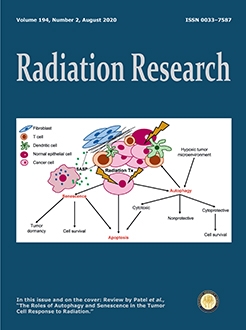
FIG. 1
In response to radiation treatment, tumor cells can upregulate both cell death and cell survival pathways. Whereas apoptotic cell death is the ideal outcome for clinical therapeutic treatment, tumor cells often enter into senescence and autophagy, largely in efforts to evade cell death. However, radiation-induced autophagy can assume different functional roles. Induction of the cytoprotective form of autophagy allows cells to evade apoptotic cell death and prolong survival; however, cytotoxic autophagy can facilitate either apoptotic and/or autophagic cell death. Finally, an alternative form of autophagy that does not appear to influence cell sensitivity to radiotherapy can occur, termed nonprotective autophagy. Senescence often occurs in parallel with autophagy, sharing a number of mechanistic regulators. Radiation-induced senescence allows cells to transiently arrest in efforts to repair damage. Subsequently, tumor cells may undergo apoptotic cell death if the extent of damage is excessive or may overcome the insult, allowing for continued survival. Senescence may also contribute to tumor dormancy, as a subset of senescent cells endure a prolonged growth arrest and regain proliferative capacity. Senescent cells produce a unique secretory phenotype (SASP), allowing for manipulation of the ECM and influencing surrounding cells in the tumor microenvironment (TME). Through the release of specific cytokines and chemokines, autophagy and senescence can play immune-modulatory effects to create either immune-promoting or immune-suppressive microenvironments, thereby contributing to overall tumor survival or clearance. Both autophagy and senescence have cell-autonomous, as well as cell-non-autonomous effects, adding to the complexity of responses and outcomes of clinical radiotherapeutics.

OCCURRENCE OF APOPTOSIS IN RESPONSE TO RADIATION
Apoptosis is a process of programmed cell death characterized by chromatin condensation, DNA fragmentation, cell shrinkage, membrane blebbing and formation of apoptotic bodies (4). While irradiated tumor cells clearly do undergo apoptotic cell death, the extent of apoptosis tends to be relatively low (5–7). Clinically relevant or even significantly higher doses of radiation induced only ∼20–30% apoptosis in several experimental tumor cell lines, including breast cancer, non-small cell lung cancer (NSCLC) and colorectal cancer (8). For instance, Rodel et al. demonstrated relative levels of apoptosis between 12% and 27% induced in response to 8 Gy in colorectal cell lines with varying radiosensitivity (7). Similarly, Qu et al. reported ∼20–25% apoptosis induction in MCF-7 breast cancer cells and A549 lung cancer cells with 8 Gy (9). In agreement with these data, previously published work performed in our laboratory demonstrated low levels of apoptosis (∼20%) induced in breast, lung, colorectal, and head and neck cell lines when exposed to fractionated radiation exposure (8). It is only at higher levels of irradiation (above 10 Gy) that apoptosis becomes a more pronounced response to radiotherapy (10). This is, of course, relevant to stereotactic radiation treatment, wherein multiple, precisely focused beams of radiation are delivered to patients to achieve higher effective doses to the tumor while minimizing damage to surrounding tissue (11–13). With regard to cancer treatment modalities, apoptosis or other forms of cell death are, of necessity, the desired outcomes; however, there are a number of survival mechanisms that cancer cells have employed to evade (apoptotic) cell death. Both autophagy and senescence can allow cancer cells to mitigate or perhaps delay the damage incurred by clinical therapeutic modalities, escape cell death and prolong survival.
OCCURRENCE OF AUTOPHAGY AND SENESCENCE IN RESPONSE TO RADIATION TREATMENT
Although the desired outcome of radiation therapy is tumor cell death by a pathway such as apoptosis, apoptosis is not obligatorily the sole or primary response to radiation treatment. While the effectiveness of clinical radiation therapy in promoting tumor shrinkage may, of necessity, ultimately involve apoptosis, possibly subsequent to mitotic catastrophe, studies in tumor cells in culture clearly indicate that a consistent and uniform initial response to radiation treatment is autophagy (14–17). Autophagy is a cellular survival mechanism to maintain homeostasis in response to stress, such as nutrient deprivation or hypoxia (18). In response to radiotherapy, autophagic machinery is upregulated to remove damage incurred by therapy. Studies by Ren et al. (19) utilizing 30 NSCLC patient tissue samples subjected to 2 Gy (a clinically relevant dose), assessed LC3 and SQSTM1/p62, markers of autophagy, by immunohistochemical staining. Of these 30 samples, 26 demonstrated significant upregulation of LC3 and downregulation of SQSTM1/p62, indictive of autophagy induction (15, 19–22).
Autophagy is generally considered to be a cytoprotective response to various forms of stress such as chemotherapy and radiotherapy (23–25), although there is accumulating evidence that autophagy can also be functionally “nonprotective” (26, 27). Nonprotective autophagy is characterized by a lack of sensitization or protection against a cellular stress or therapeutic agent when autophagy is inhibited; in such cases, autophagy is neither protective nor cytotoxic in response to the therapy (28). The current clinical utilization of autophagy inhibitors to sensitize tumor cells to radiotherapy and chemotherapy relies on the paradigm that autophagy is cytoprotective in nature; however, autophagic function can vary depending on tumor cell type, cytotoxic therapy used and treatment regimen. It is, therefore, necessary to thoroughly understand the nature of the autophagy induced to improve patient outcomes to autophagy-inhibition therapy.
Autophagy is often accompanied by senescence, at least in the case of clinically relevant doses of drugs and radiation (26, 29). Senescence has long been considered to be an irreversible form of growth arrest, although recently published studies by our group, as well as others, have shown that, while senescence is a durable form of growth arrest, tumor cells can ultimately escape from the senescent state and recover self-renewal capacity (6), possibly suggesting that senescence could be a form of tumor dormancy (30). While both autophagy and senescence often occur in parallel in response to therapeutic treatment and share a number of key regulators, such as p53 and mammalian target of rapamycin (mTOR), whether they are independent or interdependent is still not fully understood; it is feasible that a direct relationship will be evident in some systems, and not others, and will occur only with certain forms or inducers of senescence (31, 32).
RADIATION AND AUTOPHAGY
Autophagy can be considered as a “first responder”, a mechanism whereby the cell attempts to reduce the impact of cellular damage and salvage components/nutrients to avoid cell death. Our group, as well as others, have shown that autophagy is induced in response to radiation treatment and chemotherapy (8, 15, 16, 26, 33); however, the function of autophagy induced in response to these various treatment modalities is not predictable. In most of the current literature, autophagy is considered to have a cytoprotective function; consequently, inhibition of cytoprotective autophagy would be anticipated to result in radiosensitization. Early work performed by Chaachouay et al. showed that autophagy inhibition with 3-methyladenine (3-MA) and chloroquine (CQ) radiosensitized MDA-MB-231 (MDA-231) and HBL-100 breast cancer cells (16). Similarly, CQ was also shown to sensitize bladder cancer cells to radiation both in vitro and in vivo, and to promote apoptosis when autophagy inhibition was combined with radiation (34). In studies by Liang et al. examining the role of autophagy in multidrug-resistant ovarian carcinoma, radiation induced relatively low levels of apoptosis; additionally, inhibition of apoptosis with ZVAD did not significantly alter survival or cell death, further confirming that apoptosis is not the primary therapeutic response of radiation, at least in this experimental model (35). These studies also demonstrated higher basal autophagy in the multidrug-resistant phenotype SKVCR cells compared to human SKOV3 ovarian carcinoma cells, suggestive of a cytoprotective function. Moreover, inhibition of autophagy with 3-MA sensitized the multidrug-resistant cells to radiation while having only modest effects on the parental SKOV3 cells. These studies support the concept of autophagy functioning as a cytoprotective mechanism employed by the tumor cells to avert cell death, and that manipulation of these processes could hold therapeutic potential. Ko et al. also demonstrated that genetic inhibition of autophagy radiosensitizes H460 and A549 cells in vitro; however, when moved to an in vivo model of immune-competent mice, autophagy inhibition reduced responses to radiation treatment (36). This observation adds another layer of intricacy to the overall role and contributions of autophagy to tumor cell growth and host immune cell modulation.
Kuwahara et al. utilized radioresistant liver cancer cell lines, which they had previously generated, to better understand the contributions of autophagy towards radioresistance (37). These investigators demonstrated autophagy induction in response to radiation treatment in both the parental HepG2 cells and in the resistant cells (HepG2-8960-R). Furthermore, exposure to rapamycin, an mTOR inhibitor and autophagy inducer, sensitized HepG2-8960-R cells to radiation (10 Gy) but not the parental cell line. Pharmacological and genetic inhibition of autophagy reduced rather than increased sensitivity to acute radiation exposure (2 Gy), suggesting that suppression of cytotoxic autophagy could contribute to radiation resistance. However, it is generally challenging to select for radiation resistance in vitro and consequently there is relatively limited literature relating to autophagy in acquired radiation resistance.
It should be recognized that even if radiation-induced autophagy was solely cytoprotective in function, this cytoprotective form of autophagy could not uniformly represent a mechanism of resistance, since essentially all tumors undergo autophagy in response to radiation treatment and not all tumors present with radiation resistance (a degree of cell death is still observed). One possibility is that some unique (but as yet unidentified) characteristics can determine when radiation-induced cytoprotective autophagy actually confers radiation resistance, rather than simply shifting the dose-response curve for radiation sensitivity. Since the protective function of autophagy is generally considered to be a consequence of interference with apoptosis, radiation resistance could be related to this intrinsic function. A related possibility is that radiation resistance only occurs when the autophagy is durable while a transient cytoprotective outcome would not be capable of conferring actual radioresistance due to the inevitable outcome of apoptotic tumor cell death.
In our previously published studies, we identified the “nonprotective” form of autophagy. As is also the case for cytoprotective autophagy, this is a functional definition, where autophagy is induced in response to radiation treatment (or chemotherapy), but where subsequent inhibition of autophagy fails to alter radiation sensitivity (26). Eng et al. demonstrated that both pharmacological inhibition of autophagy with CQ and Lys01, and genetic inhibition by genome editing of ATG7, did not alter sensitivity to radiation (or 30 different chemotherapies) of KRAS mutant tumors in vitro and in vivo. Furthermore, they were able to show that CQ-mediated sensitization was independent of autophagy, suggesting the antiproliferative effects may be due to modulation of off-target effects (27, 38). Similarly, studies performed by Schaaf et al. demonstrated that radiosensitization effects of CQ, 3-MA and ATG7 deficiency were independent of canonical autophagy pathways and may involve effects on lysosomal degradation (39). Further analyzing these studies by Schaaf et al. (39), pharmacological inhibition with CQ and 3MA did not alter radiosensitivity in MDA-MB-231 breast tumor cells when exposed to 5.6 Gy; however, work done by Chaachouay et al. (16) showed that autophagy inhibition with similar concentrations of CQ and 3MA was sufficient to radiosensitize the same MDA-MB231 cell line exposed up to 5 Gy. These contradictory observations present a conundrum within the field when the same cell line, exposed to similar radiation doses and concentrations of the autophagy inhibitor, can produce two divergent responses, leading to opposing conclusions relating to the role of autophagy. In a seminal article by Michaud et al. (138), autophagy that was induced in colorectal cancer cells by oxaliplatin or mitoxantrone proved to be nonprotective in function, in that silencing of the autophagy gene, ATG7, failed to influence drug sensitivity in the tumor cells in vitro. However, these studies did not include radiation.
Intriguingly, work by Cechakova et al. suggested that Lys05, an autophagy inhibitor, could radiosensitize H1299 (p53-null) cells, suggesting a cytoprotective autophagic function (40). However, in fact, a close examination of data reveals that sensitization, if any, is at best modest, and unlikely to be therapeutically relevant, indicating that the autophagy is likely acting in a nonprotective fashion. This observation actually serves to confirm findings from our own laboratory where we reported radiation-induced nonprotective autophagy in the same H1299 (p53-null) cells, i.e., wherein autophagy inhibition likewise failed to alter radiosensitivity (8).
Massive tumor growth can lead to low oxygen environments, resulting in hypoxia and in nutrient deprivation, common hallmarks of solid tumors, ultimately contributing to cellular damage (41). Cells can upregulate autophagy under such stressful conditions to prevent accumulation of damaged organelles by removing and recycling cellular content (42, 43) Hypoxia often interferes with the effectiveness of anti-cancer therapies. The degree of oxygenation of a tumor can determine the cytotoxicity of radiation; thus, hypoxic environments can contribute to radioresistance (44). Under hypoxic conditions, a number of intracellular pathways are upregulated to allow tumor adaptation, such as that involving hypoxia-inducible factor 1α (HIF-1α). HIF-1α is a major sensor of hypoxic conditions and is activated to allow cells to adapt and survive under low-oxygen environments; moreover, in tumor cells, activation of this pathway acts as a survival mechanism to evade apoptotic cell death and contribute to resistance (44). Hypoxia and HIF-1α can also contribute to autophagy induction, allowing for cells to remove damaged dysfunctional organelles, such as the mitochondria (44, 45). Work by Zhong et al. demonstrated that deletion of HIF-1α in MCF-7 breast cancer cells significantly increased radiosensitivity while decreasing autophagy when irradiated (45). Similarly, Sun et al. showed that under hypoxic conditions, HIF-1α induces autophagy and reduces radiosensitivity in human colorectal cancer cell lines (46). Noman et al. showed that lung carcinoma cells under hypoxic conditions upregulated autophagy to evade CTL-mediated lysis (47). Furthermore, autophagy inhibition was sufficient to reduce tumor growth and increase apoptosis in mice transplanted with B16 melanoma cells. These studies provide evidence to support the conclusion that at least in the case of hypoxia, autophagy induction may, in fact, mediate cell autonomous effects (i.e., directly in the tumor), as well as cell non-autonomous effects, through modulation of the immune system.
Studies in our laboratory examining the functional role of autophagy in radiation sensitivity showed that inhibition of radiation-induced autophagy generally did not alter radiation sensitivity or radiation-induced ROS levels. In fact, it appears that p53 status influenced radiation sensitivity through the promotion of senescence (26). Taken together, the frequent lack of alteration in radiosensitivity exhibited when autophagy inhibition is combined with radiation suggests that autophagy frequently plays a nonprotective function and may not be the primary protective mechanism contributing to loss of radiosensitivity.
In those cases where autophagy takes on functional forms other than being protective, impairment of autophagic functions could shift the cellular fate towards unfavorable outcomes. In the case of cytoprotective autophagy, inhibition of autophagy results in increased cell death (34). In contrast, in cells where autophagy is cytotoxic or cytostatic, autophagy inhibition would be counterproductive, and permissive for tumor cell growth. In the case of cytotoxic autophagy, administration of an inhibitor would result in tumor promotion, by interfering with the original antitumorigenic actions of the autophagy in the cell. For instance, studies performed by Kim et al. demonstrated reduced radiation sensitivity in NSCLC HCC827 cells when Beclin 1, a protein required for the initiation of autophagy, was silenced, when compared to wild-type cells, suggesting that autophagy was cytotoxic in nature. Furthermore, inhibition of mTOR, ultimately resulting in autophagy induction, was sufficient to sensitize these cells to radiation (48).
As indicated above, tumor cells almost uniformly undergo autophagy in response to exogenous forms of stress such as chemotherapy and radiation treatment. Although the majority of the scientific literature tends to consider autophagy as a cytoprotective response to stress and as a mechanism of resistance, this premise is subject to a number of reservations. One is that autophagy is not uniformly cytoprotective; in fact, autophagy can exist in one of four functional forms, only one of which is protective; the other forms are cytotoxic, cytostatic and nonprotective autophagy (49). Consequently, efforts to exploit autophagy inhibition as a therapeutic strategy for radiosensitization (or chemo-sensitization) are unlikely to be successful unless all autophagic responses to radiation treatment, regardless of the tumor type, actually prove to be cytoprotective, which is highly unlikely based on our preclinical studies.
AUTOPHAGY AND TUMOR DORMANCY
Dormancy is traditionally considered to be a state of arrest wherein tumor cells cease to divide but still remain viable until appropriate environmental conditions are introduced to begin proliferation, leading to disease recurrence (50). Recently published work by Vera-Ramirez et al. demonstrated the role of autophagy in sustaining/promoting survival in dormant breast cancer cells. Autophagy inhibition via hydroxychloroquine (HCQ) exposure was sufficient to reduce cellular survival of dormant breast cancer cells and to reduce lung metastases in vivo (51). However, once these cells regained proliferative growth, the effect of HCQ was minimal, suggesting that autophagy could be important for cells in prolonging dormancy. However, it should be acknowledged that HCQ may have multiple off-target effects, raising reservations as to whether the observed effects are actually directly related to autophagy.
The potential involvement of autophagy in tumor dormancy was recently supported by published studies from our group showing that autophagy-deficient tumor cells entered the state of dormancy in the presence of chemotherapy, in vitro, and recovered earlier than autophagy-competent dormant cells (52, 53). Since autophagy is involved in the maintenance of DNA integrity such that autophagy-deficient tumor cells accumulate γ-H2AX foci and genomic damage leading to tumor progression (54), lack of autophagy could result in DNA fragmentation in surviving cells. We showed that a genetic knockdown of ATG5 resulted in the formation of multinuclear dormant tumor cells with higher DNA content in response to chemotherapy, in vitro and in vivo, leading to tumor recovery more rapidly than autophagy-competent dormant cells (52). A recent published review highlighted the paradoxical roles of autophagy in tumor dormancy and tumor progression (55), suggesting that the outcome of autophagy could depend on cellular pathways/proteins that are randomly impacted by autophagy.
RADIATION AND SENESCENCE
Senescence is a prolonged growth arrest generally associated with the induction of DNA damage and consequent signaling, involving induction of p21waf1 (and sometime p16), inhibition of cyclin-dependent kinases, dephosphorylation of Rb and the presumed formation of Rb-E2F complexes (56, 57). Senescence can furthermore be considered as an alternative cell fate which can be induced to allow cells to evade apoptotic cell death (58). Senescent cells present with a myriad of phenotypes, such as morphological changes (enlargement and flattening), expression of a pH6-dependent beta-galactosidase activity, secretion of chemokines and cytokines that encompass the senescence-associated secretory phenotype (SASP), and heterochromatic foci appearance (59–61). Previously published studies have quite conclusively demonstrated growth arrest characteristic of senescence in response to radiation treatment (62–66). Cui et al. showed that a dose of 4 Gy to cervical cancer cells induced only 16% apoptosis but did induce a long-lasting G2/M-phase arrest (67). Recently reported work from our research group demonstrated that H460 NSCLC cells also exhibit senescence induction in response to radiation treatment and arrest at the G2/M phase (26). In agreement, Luo et al. showed that a 6 Gy dose did not induce significant apoptosis in A549 and H460 cells, but rather induced a premature senescence indicated by increased SA-β-gal staining. Furthermore, knockdown of p53 inhibited radiation-induced senescence, while restoration of p53 expression sensitized cells to radiation and induced senescence (68). Studies performed by Roberson et al. demonstrated induction of accelerated senescence in response to camptothecin, a DNA-damaging agent, in p53-null H2199 human lung cancer cells. Of interest was that these investigators demonstrated that a subset of cells were able to escape the therapy-induced senescence and these cells resembled parental cells while still exhibiting SA-β-gal activity (69). In recently reported work, our group was able to demonstrate that radiation induced a transient growth arrest characterized as senescence, followed by proliferative recovery in NSCLC cells. Moreover, radiation-induced senescence was greater in p53wt H460 NSCLC cells, which were more sensitive to radiation treatment than the p53-null H460 cells (26). These data suggest that p53, an essential tumor suppressor protein commonly mutated in many cancer types, plays a role in mediating senescence induction in response to radiation-related damage, and furthermore, that differential induction of senescence may be a primary contributor to altered radiation sensitivity in various cancers (26, 70).
Aside from its tumor-suppressive functions, senescence may also present as a model of tumor dormancy (30). Damage incurred by the cells from radiation and chemotherapy can induce senescent growth arrest as the cell attempts to repair the damaged DNA. Radiation has been shown to induce a temporary period of growth arrest, followed by a phase of proliferative recovery (26, 69, 71) that could theoretically contribute to disease recurrence.
One limitation to efforts to fully understand the role and contributions of therapy-induced senescence in radiosensitivity is the absence of specific pharmacologic or genetic approaches to silence the senescence response. Nevertheless, our recent evidence of outgrowth/escape from senescence argues for the likelihood that senescence, like autophagy, may be a cytoprotective response that allows the tumor cells to escape elimination by radiation. The prolonged and sustained growth inhibition may be permissive for the ultimate regrowth of the tumor cells and the consequent disease recurrence. Given this possibility, coupled with evidence that the senescence-associated secretory phenotype (SASP) may also promote tumor growth, the recent identification of agents with “senolytic” properties, which promote apoptosis selectively in senescent cells, opens up the possibility of developing a therapeutic strategy for elimination of the residual surviving tumor cells (72, 73).
Work by Yosef et al. (74) demonstrated that human fibroblasts induced into senescence by radiation upregulated anti-apoptotic proteins, BCL-W and BCL-XL, in vivo. Targeting these proteins using a small-molecule inhibitor, ABT-737, was sufficient to eliminate these senescent cells (74). Similarly, Samaraweera et al. (75) showed senescence induction and SASP secretion in both NSCLC cells and head and neck squamous cell carcinoma (HNSCC) cells when exposed to cisplatin or taxanes. Furthermore, administration of panobinostat, an FDC-approved histone deacetylase inhibitor (HDACi), after chemotherapy, suppressed proliferation and induced cell death in both cancer cell types (75). While there are many agents that have been proposed to have properties of senolytics (72, 76), not all of these compounds have been shown to be universally effective; consequently, further work will be required to generate clearer insights as to exactly how these agents act as “senolytics” and why particular agents are effective under certain experimental conditions but not others. Taken together, selectively targeting senescent cells while they are dormant and before they begin to regain proliferative recovery may serve as a therapeutic benefit and prolong patient survival, as well as increasing the delay before disease recurrence.
AUTOPHAGY AND SENESCENCE
Autophagy and senescence are both induced in response to radiation treatment, which may in part be due to the overlapping pathways and shared regulators between these two processes.
Reactive Oxygen Species
A key mechanism by which radiation treatment exerts its indirect cytotoxic effects is through ROS generation. Increased ROS levels can be detrimental to the intracellular environment, resulting in mitochondrial dysfunction, genomic instability and misfolded proteins (77). To mitigate the ROS-mediated stress, autophagy is induced to remove and turn over damaged organelles and proteins (78). Chen et al. demonstrated ROS elevation by low-dose ionizing irradiation after high-dose irradiation promoted autophagy and induction of radioresistance in A549 NSCLC cells (79). Furthermore, treatment with N-acetyl-l-cysteine (NAC), an ROS scavenger, was sufficient to block autophagy and the associated radioresistance. Oxidative stress as well as the direct radiation effects can cause substantial DNA damage within a cell, which may induce a senescent cell cycle growth arrest as the cell attempts to repair the damaged DNA (80). Luo et al. showed that resveratrol enhanced radiosensitivity of H460 and A549 NSCLC cells by ROS-mediated DNA damage which induced a premature senescence (64, 81). ROS-induced DNA damage upregulates and activates a number of important cellular regulators, including p53, which in turn increases p21, a key cell cycle regulator in mediating senescence (82).
TP53
TP53 is an essential tumor suppressor gene, which is commonly aberrant or dysfunctional in multiple tumor cell types. p53 regulates a vast number of cellular processes, including but not limited to apoptosis, autophagy and senescence. p53 mediates the transcription of a number of cell cycle inhibitors, including p21waf1 and p16, which interfere with the interaction between cyclins and cyclin-dependent kinases that induce cell cycle arrest (83). Luo et al. demonstrated that p53 activation using Nutlin-3a radiosensitized H1299 (p53-null) cells by activating p53-p21waf1 pathways and inducing cellular senescence (68). Furthermore, depending on p53 localization, this protein can also mediate autophagy. p53 can activate autophagy by upstream inhibition of mammalian target of rapamycin (mTOR), through direct means such as generation of damage-related autophagy modulators (DRAM), as well as regulating the transcription of key autophagy-related genes (84–86).
Mammalian Target of Rapamycin (mTOR)
Mammalian target of rapamycin (mTOR) is an important regulator of both senescence and autophagy. mTOR prevents activation of autophagy initialization; thus, mTOR inhibition has been shown to upregulate autophagy (87). Cao et al. showed that addition of RAD001, a pharmacological mTOR inhibitor, enhanced radiosensitivity of prostate cancer cell lines, which was associated with increased autophagy (88). Studies by Nam et al. demonstrated autophagy activation in response to mTOR inhibition in glioma, lung, colorectal and breast cancer cell lines when exposed to radiation; furthermore, mTOR blockade (which promotes autophagy) resulted in premature senescence and restoration of radiosensitivity (89). Seminal work by Narita et al. showed that mTOR and autophagic machinery may be important in SASP processing during senescence (90, 91). The authors observed a specialized compartment, which they termed the TOR-autophagy spatial coupling compartment (TSACC), where products of cellular catabolic processes, such as autophagic degradation, could feed into cellular anabolic processes, to promote protein synthesis. Disruption of mTOR localization to TSACC was shown to inhibit interleukin-6/8 synthesis in Ras-induced senescence, suggesting that autophagy may play a role in SASP generation, which can reinforce the senescent phenotype.
Recently published studies from our laboratory utilizing multiple cytotoxic therapies and multiple cell lines demonstrated senescence induction and proliferative recovery independent of autophagy when the autophagy is “nonprotective” in function (92). Furthermore, we observed that autophagy inhibition did not alter the extent of senescence induction or recovery in HCT116 colorectal carcinoma cells exposed to 4 Gy. Given that both autophagy and senescence are induced in parallel in response to radiation treatment, and often occur in conjunction, it has been asserted that senescence is dependent on prior autophagy. However, a careful analysis of the literature suggests that while autophagy inhibition can delay the occurrence of senescence, it is unlikely to alter the extent of senescence induction; that is, the promotion of autophagy may merely accelerate the senescence response, but senescence is not autophagy-dependent (71, 93, 94).
BYSTANDER AND ABSCOPAL EFFECT/INVOLVEMENT OF THE IMMUNE SYSTEM IN THE RESPONSE TO RADIATION TREATMENT/DAMPS/SENESCENCE
Autophagy and senescence both can exhibit immune-modulatory effects, both endogenously and exogenously. Endogenously, autophagy is essential for T-cell activation and differentiation in response to environmental insults. Dysregulation of autophagy and an increase of senescence during aging of T cells results in a compromised immune response to pathogens in the elderly (95). Exogenously, through SASP secretion, senescent cells can regulate immune infiltration and clearance of tumor cells, while autophagy contributes to immune cell function and cytokine production (72, 96).
Senescence-Associated Secretory Phenotype
A key characteristic of senescent cells is the senescence-associated secretory phenotype (SASP), which encompasses a unique secretion profile that includes cytokines, matrix metalloproteases (MMPs), growth factors and several other soluble regulators (97). A number of these factors can alter the tumor microenvironment (TME) or promote tumor growth. Additionally, several of these cytokines and soluble factors participate in wound-healing processes that modulate extracellular matrix (ECM) remodeling, tissue repair, surrounding cells in the TME, as well as regulate immune infiltration (98). MMPs and other proteases help to remodel the ECM around tumor cells and can promote tumor migration (99, 100). Moreover, senescent cells secrete a number of pro-inflammatory cytokines and chemokines, such as IL-6 and IL-8, which can play a role in promoting tumor growth and migration. Ortiz-Montero et al. demonstrated that when MCF-7 cells were treated with conditioned media from senescent cells, IL-6 or IL-8, markers of endothelial-to-mesenchymal transition (EMT) were upregulated, and promoted tumor cell migration and invasion (101). Both of these inflammatory factors can modulate the activation of the NF-κB pathway, resulting in an increase in transcription of anti-apoptotic proteins and promoting tumor growth (102). In contrast, secretion of pro-inflammatory cytokines can promote immune infiltration, and depending on the type of immune infiltrates, can promote or inhibit tumor growth. Published studies by Meng et al. utilized radiation and the PARP inhibitor, veliparib, to promote premature senescence in B16SIY melanoma cells (103). When these senescent cells were isolated and injected into C57BL/6 mice, a number of cytokines were upregulated and an increased CD8+ T-cell proliferation was observed in coculture studies. Furthermore, in tumor-bearing mice, vaccination with senescent cells followed by irradiation was sufficient to elicit immune responses and eliminate established tumors. As exemplified, cellular senescence can play a dual role in tumor development itself, and it remains to be determined whether these soluble factors produced by senescent cells promote or antagonize tumorigenesis (104).
Bystander and Abscopal Effect
Given the functional interplay between SASP secretion and immune modulation, it is important to acknowledge the potential contributions of senescence to the abscopal effect. Depending on the soluble factors released by senescent cells, recruitment and promotion of the clonal expansion of a myriad of immune cells, ranging both dichotomies of pro-to anti-tumorigenic immune cells, can occur (72, 105, 106). Work by Xue et al. demonstrated that restoration of p53-rescued senescence induction, activation of the innate immune system and subsequent clearance of tumor cells (107). Furthermore, it has been demonstrated that the SASP released by senescent cells promote a bystander effect both in vitro and in vivo (108, 109), in which secretion can promote premature senescence in surrounding cells, possibly enhancing responses. Ruhland et al., showed that SASP secretion by senescent stromal cells can increase localization and activation of myeloid suppressor cells, creating an immunosuppressive, tumor-permissive environment (110). Reiterating the duality in function (104), SASP secretion can promote either an immune-suppressive effect to promote tumor growth (111) or immune-activating TME, promoting anti-tumor effects, immediately surrounding the tumor; however, whether this may contribute to distal tumor clearance has yet to be determined.
With increasing utilization of immunotherapies as an additive to the current treatment regimen of various cancer types, better understanding and manipulation of the abscopal effect could be of benefit, aiding to activate immune responses and increasing immune clearance in otherwise “cold” tumors.
Tumor immune surveillance could keep nascent transformed cells in check and inhibit tumor formation (112); however, “cold” tumors or weakly immunogenic tumors could escape tumor immune surveillance. Tumors that do not contain infiltrating lymphocytes are termed non-inflamed or immunogenically “cold” tumors, while those with predominant immune cell infiltrates are termed inflamed or immunologically “hot” tumors. Both “cold” tumors and weakly immunogenic tumors could be turned into “hot” or highly immunogenic tumors due to radiation exposure, which could result in inflammation and immunogenic cell death (ICD). Subsequently, tumors undergoing ICD and expressing damage-associated molecular pattern (DAMP) can be detected by antigen-presenting cells (APCs) to induce antitumor immune responses (113). This phenomenon has been characterized as abscopal effects, such that direct irradiation of tumor cells on one side of an experimental animal can result in shrinkage of the tumor on the contralateral site (114) or produce a durable anti-tumor immune response and inhibit tumor progression (115). This abscopal or bystander effect has also occasionally been observed in patients where tumors distant from the original site of irradiation have also undergone reduction (116). It is understood that this is likely to be the consequence of the establishment of anti-tumor immune responses against shared tumor antigens expressed on irradiated tumor and nonirradiated tumor cells. Paradoxically, tumor-derived cytokines/chemokines could increase myeloid-derived suppressor cells (MDSCs) or Tregs, resulting in the inhibition of the anti-tumor immune responses. To this end, the contribution(s) of tumor cell autophagy or senescence in determining the outcome of immunogenic radiation therapy remains elusive.
What has not been extensively explored is whether autophagy or senescence is involved in the abscopal or bystander effects. It is well established that autophagy plays a key role in the phagocytic process, regulating the processing and secretion of inflammatory cytokines, the processing and loading of antigenic peptides, and regulating activation of adaptive immune cells (117–121). Moreover, autophagy contributes to the modulation of ICD. In their published studies, Ko et al. were able to show reduced radiosensitivity in autophagy-deficient cells in mouse tumor xenografts in wild-type immune-competent mice (36). This alteration in radiosensitivity could be rescued by recapitulating extracellular ATP in mice transplanted with ATG knockdown cells when compared to shControl transplanted mice treated with extracellular ATP and radiation, suggesting that autophagy contributes to the generation/release of ATP, a well-established DAMP recognized by the immune system. These effects were not evident in parallel experiments performed with mouse tumor xenografts in immunodeficient animals. Collectively, these experiments establish that autophagy can contribute to radiation sensitivity through its cell non-autonomous effects by modulating immune responses.
ARE PATIENT-DERIVED TUMOR XENOGRAFTS AN APPROPRIATE MODEL FOR STUDIES OF RADIATION SENSITIVITY?
As evident in the previous section, cell non-autonomous effects of autophagy and senescence, tumor microenvironment and immune cell regulation all appear to play a role in modulating sensitivity to cancer therapy; thus, the question arises whether current pre-clinical models will reflect clinical outcomes and the translational potential of novel pre-clinical therapeutics. Patient-derived xenografts (PDX) are currently considered the “gold standard” in modeling tumors and their microenvironment, in part because they can reflect the heterogeneity of tumor populations, and capture various host cell types (such as endothelial cells and fibroblasts) that compose the tumor microenvironment (122–124). These have become the preferred model to study drug sensitivity in tumors as well as chemotherapy and radiation resistance (125–127). PDX models are generally implanted in immunodeficient animals to prevent their rejection. When assessing effects of radiation, PDX models have been preferred to syngeneic ones, as rodents and humans differ significantly in processes governing response to radiation treatment (126). With the goal of simulating human disease response, the primary use of PDXs is justified. In one pioneering study, PDX models of glioblastoma were effectively used to determine whether amplification of EGFR in tumors was related to their radiation sensitivity (128). Further, mouse-specific radiation delivery techniques have been developed that closely mirror systems used clinically. In addition, tools like micro-CT have helped to evaluate the response of murine tumors to radiation (129). It should be noted, however, that for radiation therapy studies, athymic nude mice are the most reliable hosts for PDXs. Other immunodeficient mice are more radiation-sensitive due to altered DNA repair mechanisms resulting in significant changes to the tumor microenvironment (130). As an alternative to using murine hosts, PDXs can also be cultured using 3D model systems to study effects of radiation. This significantly reduces the costs associated with in vivo experiments but has still been shown to produce different data from that acquired in mono-layer culture experiments (130).
Despite the many benefits PDX models provide, their lack of an intact immune system results in limitations, especially as immunotherapies emerge as an additional pillar of therapy and are now frequently being incorporated into treatment regimens. Multiple studies have revealed the effectiveness of combination immunotherapy and stereotactic radiation in treating a variety of mouse lesions, including brain, colon and lung (131–133). Although both syngeneic and genetically-engineered mouse models (GEMM) retain functional immune systems, the many unique aspects of GEMMs are now being explored. These models, which allow for the induction of specific mutations that drive tumorigenesis, have shown utility in studying radiation therapy. Specifically, Cre recombinase and FLP-FRT systems allow for the manipulation of multiple genes to study how these differences affect response to radiation treatment.
In GEMM's of non-small cell lung cancer, genetic differences in phenotypically identical cells were shown to be related to a growth delay after irradiation (129). Although PDXs provide an effective platform for mimicking human disease response to radiation treatment, where combination immunotherapies or an understanding of specific tumorigenic mutations is concerned, syngeneic or GEMM models should be utilized for current studies. Once the baseline utility of immune-compromised mice that have been implanted with human immune cells, “humanized NOD scid gamma mice”, has further progressed, these models may be ideal to use for radiation studies.
AUTOPHAGY INHIBITION IN CLINICAL TRIALS OF RADIATION RESPONSE
While there are several pharmacological autophagy inhibitors, the only clinically approved inhibitors are chloroquine and hydroxychloroquine (HCQ) (134). HCQ is an FDA-approved drug for the treatment and prevention of malaria. HCQ is a lysomotropic agent which can also be utilized to block autophagy through its ability to inhibit the fusion of the autophagosome with the lysosome (134). In 2007, phase I/II clinical trials were performed in which HCQ was combined with temozolomide and radiotherapy in patients diagnosed with glioblastoma multiforme (22). Electron microscopy was utilized to show that radiation induced autophagic vesicle formation in patients; however, treatment with HCQ, even at relatively high concentrations, did not sufficiently inhibit autophagy. Higher doses of HCQ are required to adequately inhibit radiation-induced autophagy in patients; however, this may lead to unwanted toxicities (22, 134). Subsequent phase I/II clinical trials were also initiated in 2013 by Susan Short's group (Brazil et al.; University of Leeds, Leeds, UK) in which the combinatorial effects of lower doses of HCQ and short-course radiotherapy were examined in aged patients with high-grade glioma (HGG) (135). HCQ in combination with radiation did not improve patient outcome compared to radiation alone, and in fact, even at the lower doses of HCQ, grade 3–5 toxicities occurred more frequently in patients who received combination treatment compared to radiation alone. Several other clinical trials were initiated to study the effects of autophagy inhibition on chemo- and radiosensitivity; however, these have provided inconsistent results. HCQ as a monotherapy in patients with metastatic pancreatic adenocarcinoma provided inconsistent autophagy inhibition and poor therapeutic efficacy in a phase II trial (136). A clinical trial of HCQ treatment combined with docetaxel in patients with metastatic prostate cancer was terminated early due to a lack of improved efficacy (identifier: NCT00786682). However, a number of clinical trials have been launched utilizing higher doses of HCQ in combination with chemotherapeutics, such as bortezomib, which may show more promising results (137) (identifiers: NCT00568880, NCT01206530, NCT01506973).
SUMMARY
The premise that autophagy confers resistance to various treatment modalities has been the basis for ongoing clinical trials combining chemotherapy (or radiation treatment) with the only FDA-approved agents that have been established to inhibit autophagy (at least in vitro, the antimalarial drugs, chloroquine and hydroxychloroquine). However, there are a number of conceptual and experimental reservations relating to these clinical trials strategies. One is that, as we and others have shown in multiple published studies, autophagy is not uniformly cytoprotective, often exhibiting cytotoxic and nonprotective functions. The other is that autophagy is unlikely to actually function as a mechanism of radiation resistance if, in fact, autophagy routinely occurs in response to radiation treatment in every tumor studied to date. From a directly clinical perspective, it is highly uncertain whether chloroquine and hydroxychloroquine can achieve levels in the circulation and the tumor to actually inhibit autophagy to the extent that radiosensitization might occur. Finally, if and when more efficacious autophagy inhibitors are identified, it is necessary to acknowledge that autophagy also provides homeostatic regulation in normal tissues such as the central nervous system, and therefore autophagy inhibition might represent a double-edged sword that would induce unanticipated and undesirable (intolerable) toxicities.
While autophagy plays a role in initial sensitivity to radiation in tumor cells, accumulating literature has supported the induction of a prolonged growth arrest, characteristic of senescence, as a response to radiation treatment. Furthermore, a subset of these senescent cells is capable of regaining proliferative capacity, a possible contributor to tumor dormancy and disease recurrence. Consequently, senescent growth arrest may provide a significant contribution to radiation resistance and disease reemergence. Although the implications of senescence cannot be truly resolved due to a lack of effective inhibitors of senescence induction, a novel class of drugs has arisen that allow us to further probe this question. Senolytics selectively induce cell death in senescent cells, providing further insights relating to radiation-induced senescence, as well as a novel class of therapeutics to add to the arsenal of cancer chemotherapy. Moreover, given the fact that autophagy and senescence often occur in tandem, it is also important to further consider the therapeutic implications of incorporating autophagy inhibitors and senolytics in combination with radiotherapy as a means of eliminating residual tumor cells, which have evaded cell death.
ACKNOWLEDGMENTS
Work from the laboratory of DG was supported by the Office of the Assistant Secretary of Defense for Health Affairs through the Breast Cancer Research Program (grant no. W81XWH-14-1-0088), National Institutes of Health (grant no. 5T32DA007027 and RO1 CA 239706) and Massey Cancer Support Grant (grant no. P30 CA016059). Additional support was provided by Susan G. Komen® (to JCH, grant no. CCR19608826).