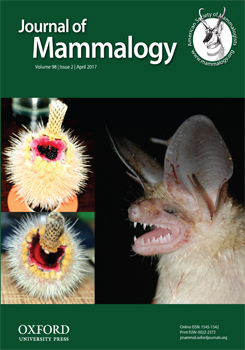
Environmental factors, including exposure to anthropogenic factors such as endocrine disruptors, can affect the luminal fluid of the epididymides in which sperm reside during maturation, causing male reproductive dysfunction. We describe and compare epididymal morphology, histomorphometry, and proteomes of Calomys tener and Necromys lasiurus, 2 species of South American sigmodontine rodent whose reproductive biology has been little studied. Five C. tener and 6 N. lasiurus were collected in a protected area of Atlantic Forest (Minas Gerais State, Brazil), where exposure to anthropogenic influences should be minimal. The left epididymis was processed for histological analysis under light microscopy, and the right was used to assess protein expression using shotgun proteomics. Calomys tener presented higher mean values for luminal and tubular diameters than N. lasiurus in the caput region. We observed similar morphologies and relative frequencies in the epididymal epithelium of principal, basal, and clear cells in the 2 species. Shotgun proteomics detected 58 and 64 proteins in 1 or more epididymal regions of C. tener and N. lasiurus, respectively. Aldose reductase, superoxide dismutase Cu-Zn, carboxylesterase 5A, and clusterin were only detected in the epididymis of N. lasiurus. The epididymides of C. tener and N. lasiurus differed in both histomorphometry and protein expression, suggesting that describing the epididymis in closely related species may provide a complementary tool for taxonomic studies. Knowledge of epididymal histophysiology helps establish a foundation for better understanding of the reproductive biology of these rodents, and our data from a protected area create a baseline for studies investigating the effects of environmental endocrine disruptors on functionality of the epididymal epithelium.
BioOne.org will be down briefly for maintenance on 17 December 2024 between 18:00-22:00 Pacific Time US. We apologize for any inconvenience.
How to translate text using browser tools
1 February 2017
Proteomes and morphological features of Calomys tener and Necromys lasiurus (Cricetidae, Sigmodontinae) epididymides
Tatiana Prata Menezes,
Mariana Moraes de Castro,
Juliana Alves do Vale,
Arlindo A. A. Moura,
Gisele Lessa,
Mariana Machado-Neves
ACCESS THE FULL ARTICLE

Journal of Mammalogy
Vol. 98 • No. 2
April 2017
Vol. 98 • No. 2
April 2017


