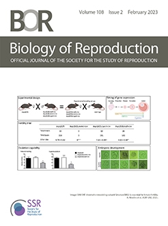
Uterine fluid plays important roles in supporting early pregnancy events and its timely absorption is critical for embryo implantation. In mice, its volume is maximum on day 0.5 post-coitum (D0.5) and approaches minimum upon embryo attachment ∼D4.0. Its secretion and absorption in ovariectomized rodents were shown to be promoted by estrogen and progesterone (P4), respectively. The temporal mechanisms in preimplantation uterine fluid absorption remain to be elucidated. We have established an approach using intraluminally injected Alexa Fluor™ 488 Hydrazide (AH) in preimplantation control (RhoAf/f) and P4-deficient RhoAf/fPgrCre/+ mice. In control mice, bulk entry (seen as smeared cellular staining) via uterine luminal epithelium (LE) decreases from D0.5 to D3.5. In P4-deficient RhoAf/fPgrCre/+ mice, bulk entry on D0.5 and D3.5 is impaired. Exogenous P4 treatment on D1.5 and D2.5 increases bulk entry in D3.5 P4-deficient RhoAf/fPgrCre/+ LE, while progesterone receptor (PR) antagonist RU486 treatment on D1.5 and D2.5 diminishes bulk entry in D3.5 control LE. The abundance of autofluorescent apical fine dots, presumptively endocytic vesicles to reflect endocytosis, in the LE cells is generally increased from D0.5 to D3.5 but its regulation by exogenous P4 or RU486 is not obvious under our experimental setting. In the glandular epithelium (GE), bulk entry is rarely observed and green cellular dots do not show any consistent differences among all the investigated conditions. This study demonstrates the dominant role of LE but not GE, the temporal mechanisms of bulk entry and endocytosis in the LE, and the inhibitory effects of P4-deficiency and RU486 on bulk entry in the LE in preimplantation uterine fluid absorption.
Summary Sentence
Visualization of the temporal mechanisms of uterine fluid absorption via bulk entry and endocytosis during early pregnancy provides novel insights into cellular and molecular mechanisms in establishing uterine receptivity for embryo implantation.
Graphical Abstract


