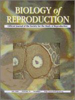
Although mammals produce either sperm or eggs depending on their sex, we found oocytes in the testes of newborn MRL/MpJ male mice. In the present study, we report the morphological characteristics of testicular oocytes, the postnatal change of oocyte number per testis, and the expression of a few oocyte-specific genes in the testes of MRL/MpJ mice. The testicular oocytes had a diameter of 50–70 μm and were surrounded by zonae pellucidae, which were observed between oocytes and follicular epithelial cells. Ultrastructurally, the testicular oocytes contained numerous microvilli and cortical granules, receiving cytoplasmic projections from follicular epithelial cells. The testicular oocytes appeared as early as at birth, and the largest number was found on Day 14. The testicular oocytes were detected in only MRL strains and B6MRLF1, but not in C57BL/6, C3H/He, BALB/c, DBA/2, A/J, and MRLB6F1. The expression of the oocyte-specific genes Zp1, Zp2, Zp3, and Omt2a was detected in testes from MRL/MpJ mice. These results suggest that newborn male MRL/MpJ mice with XY chromosomes can produce oocytes in their testes and that one of the genes causing this exists on the Y chromosome.
How to translate text using browser tools
1 July 2008
Oocytes in Newborn MRL Mouse Testes
Saori Otsuka,
Akihiro Konno,
Yoshiharu Hashimoto,
Nobuya Sasaki,
Daiji Endoh,
Yasuhiro Kon
ACCESS THE FULL ARTICLE

Biology of Reproduction
Vol. 79 • No. 1
July 2008
Vol. 79 • No. 1
July 2008
follicular development
meiosis
Mouse
MRL/MpJ
oocyte development
ovary
sex differentiation


