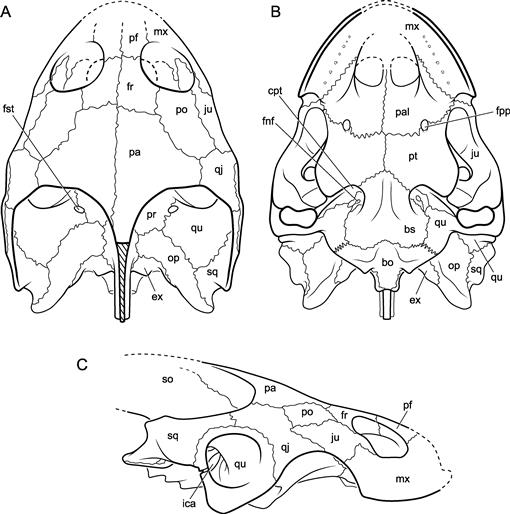
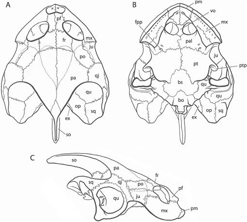
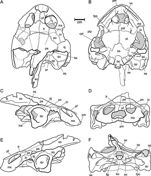
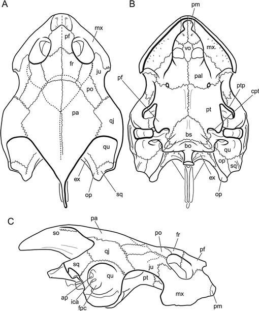
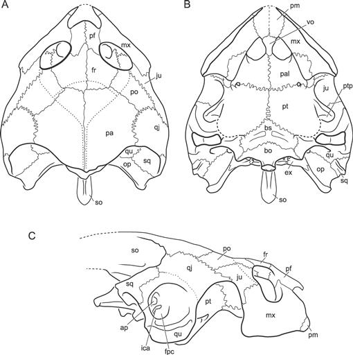
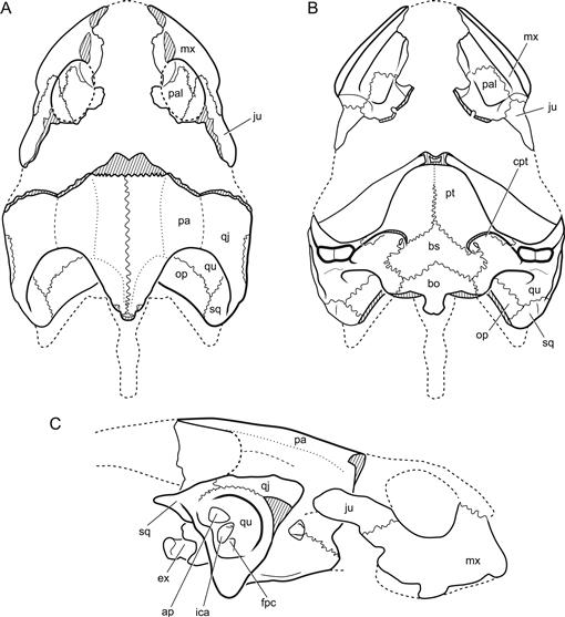
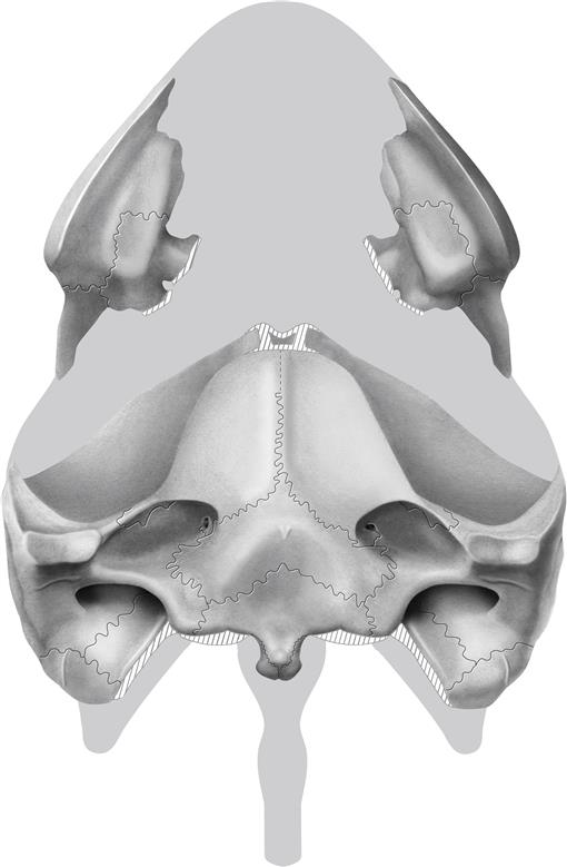
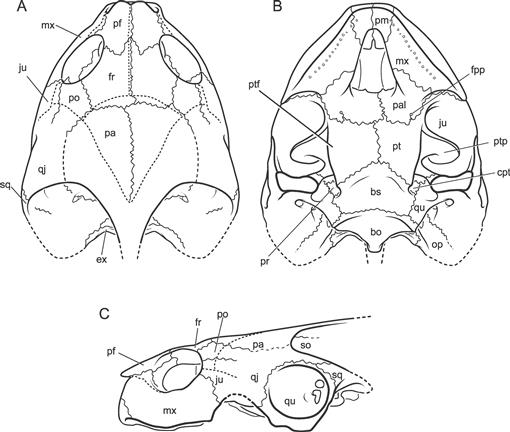
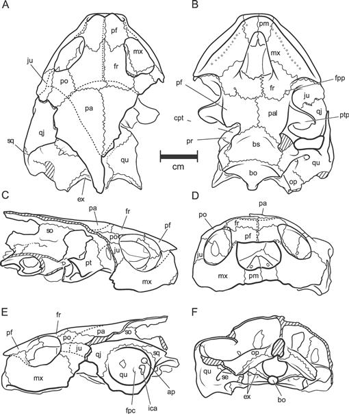
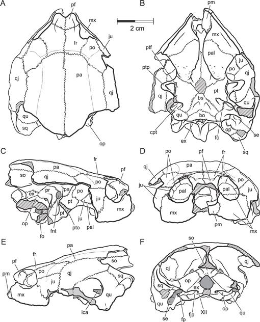
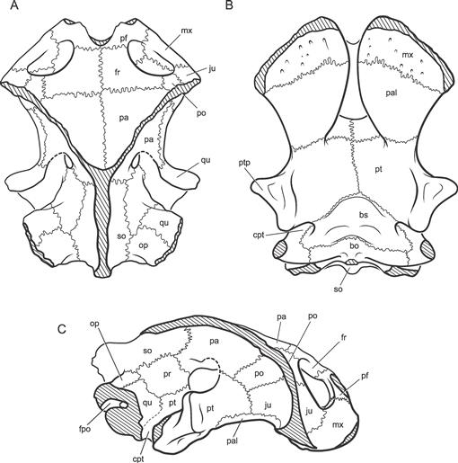
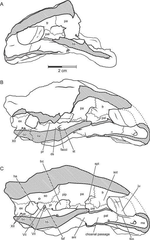
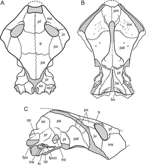
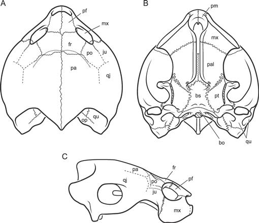
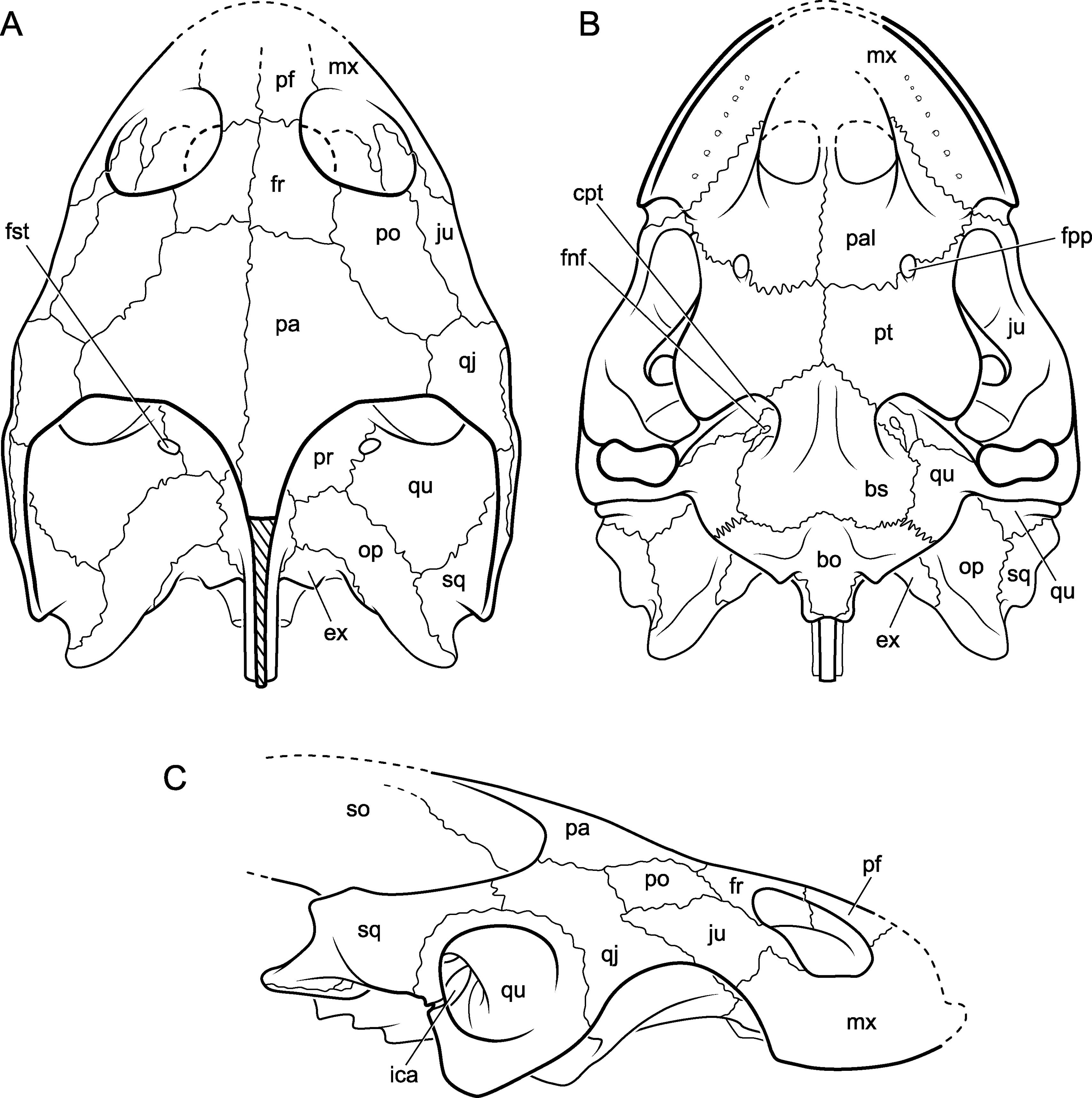
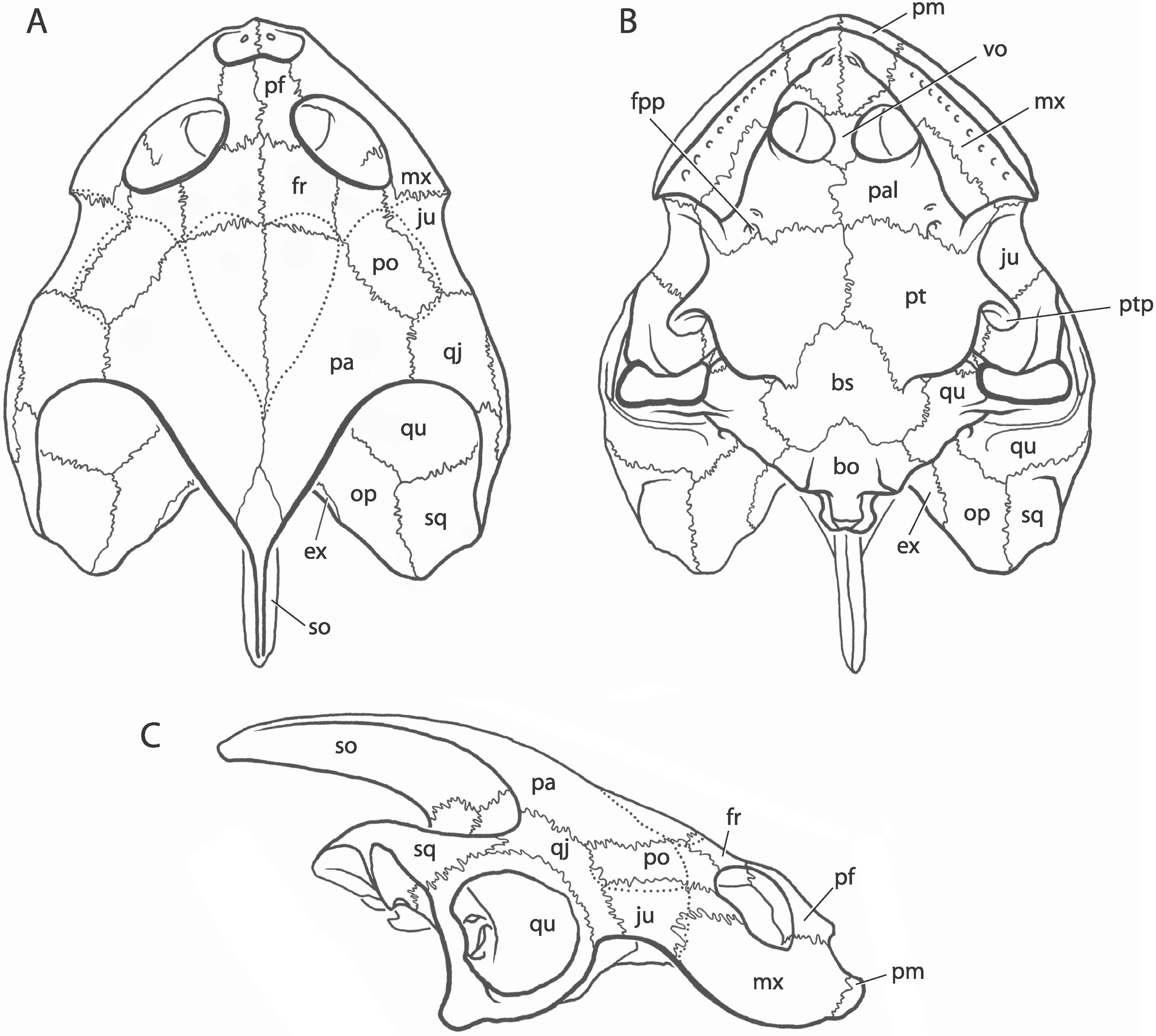
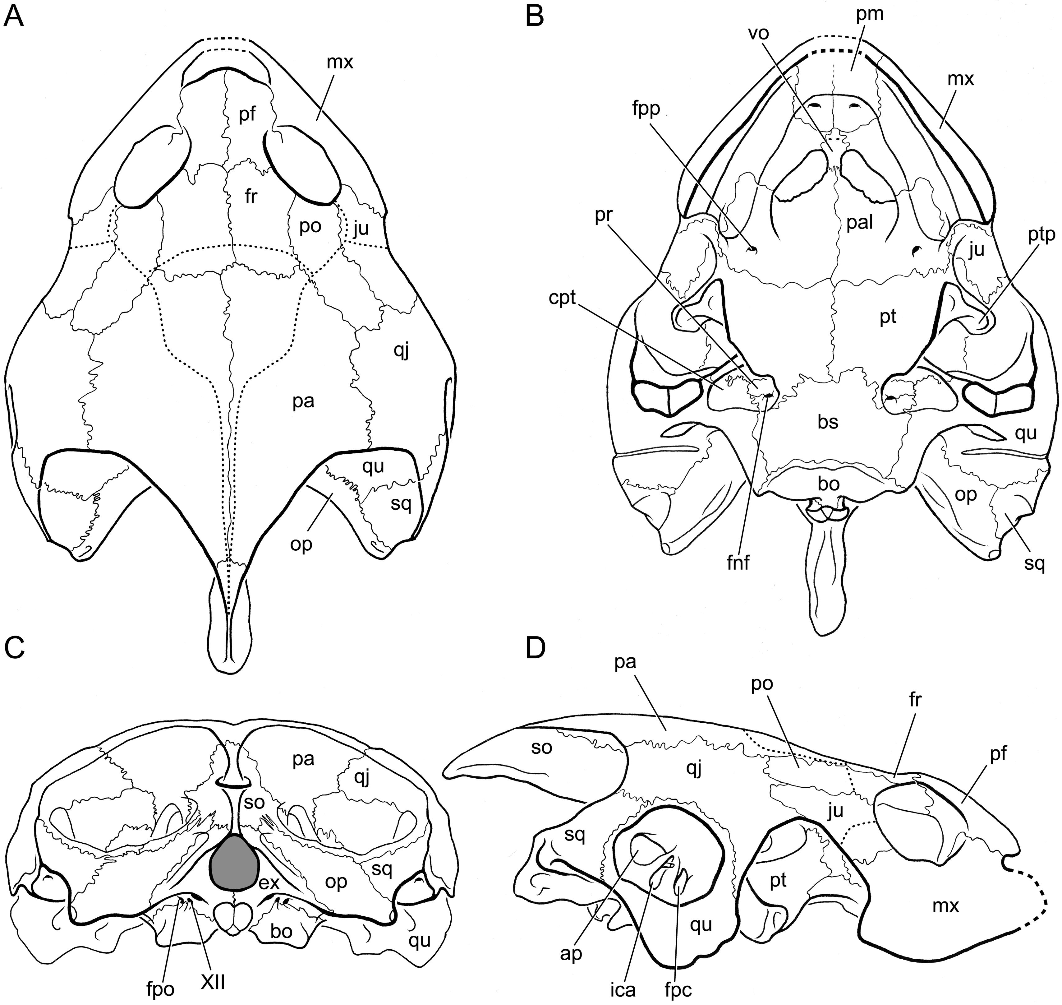
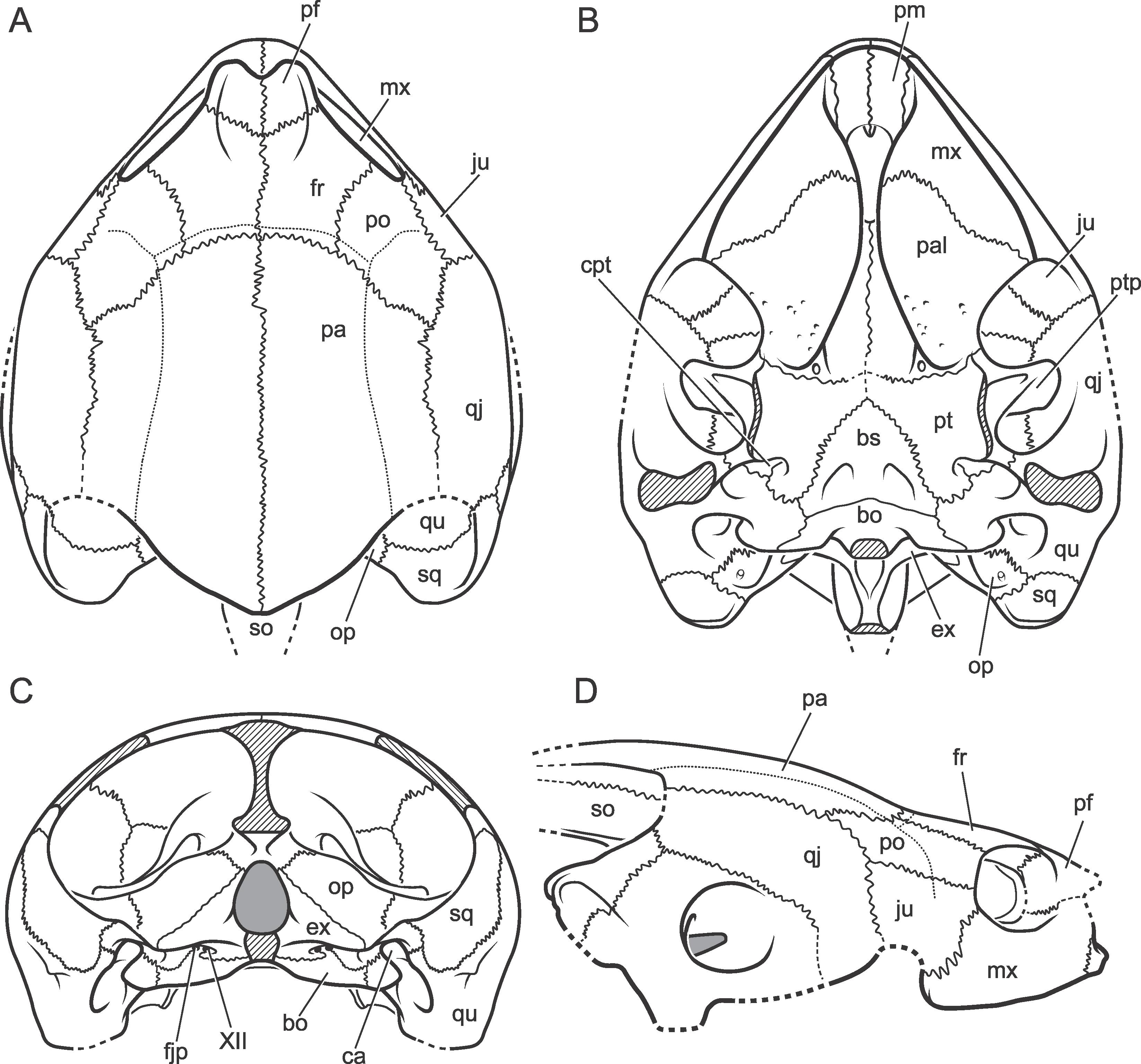
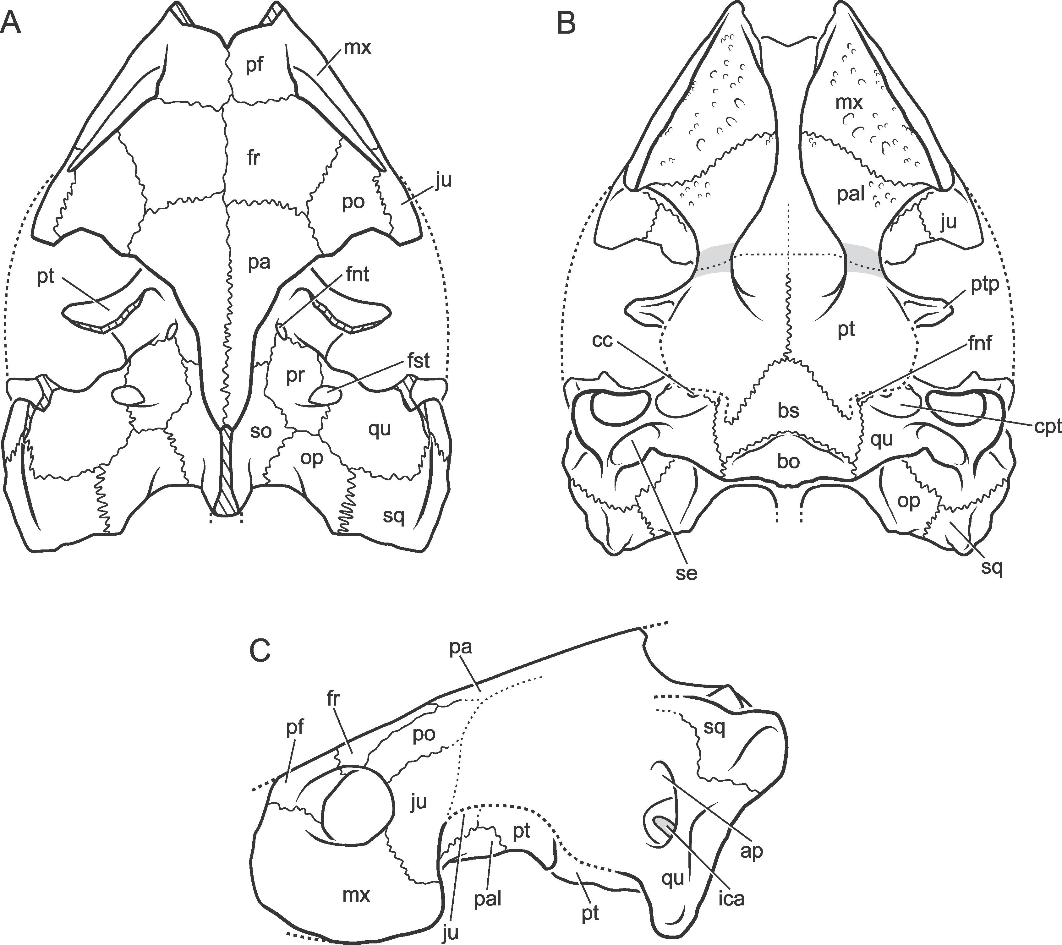
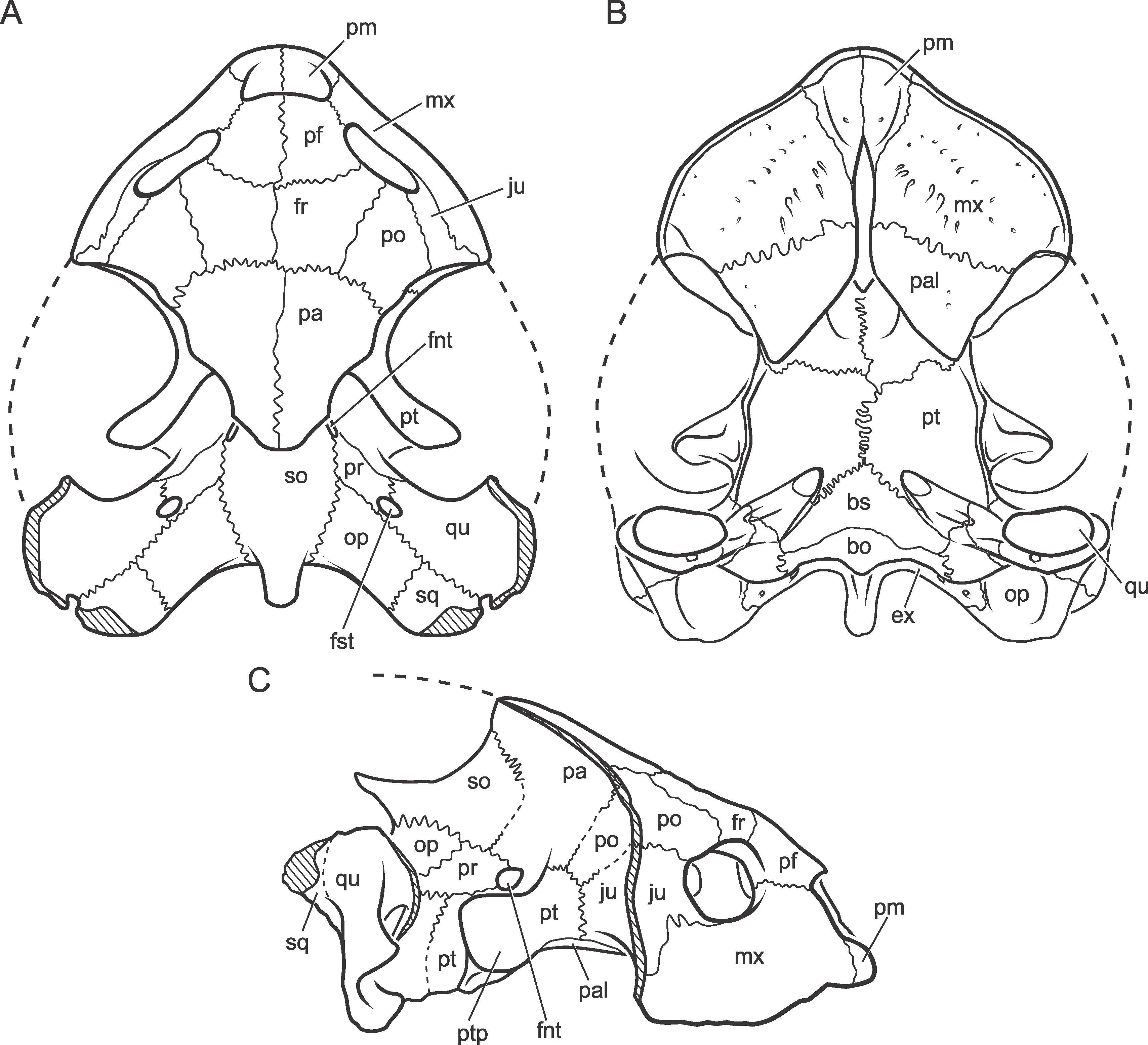
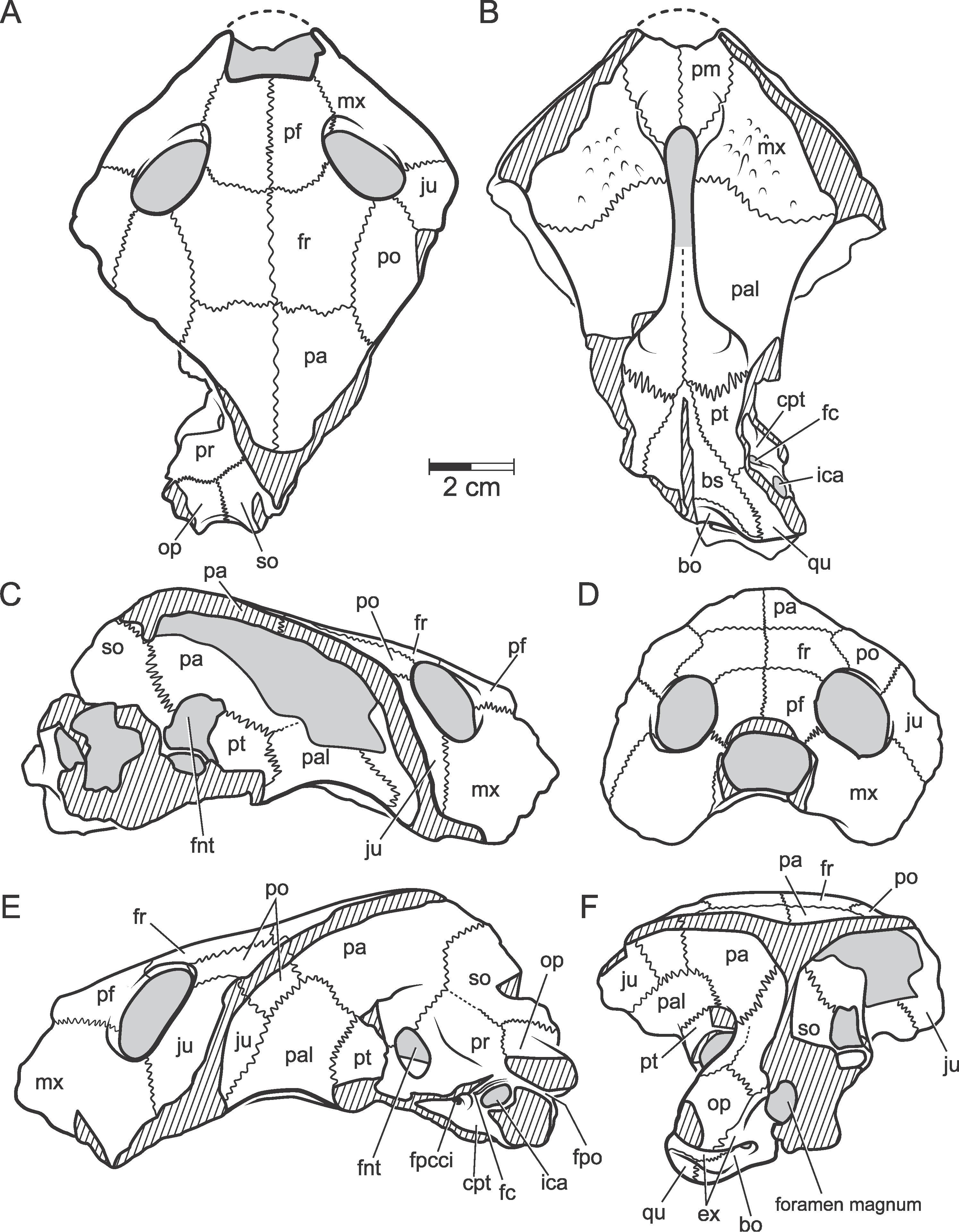
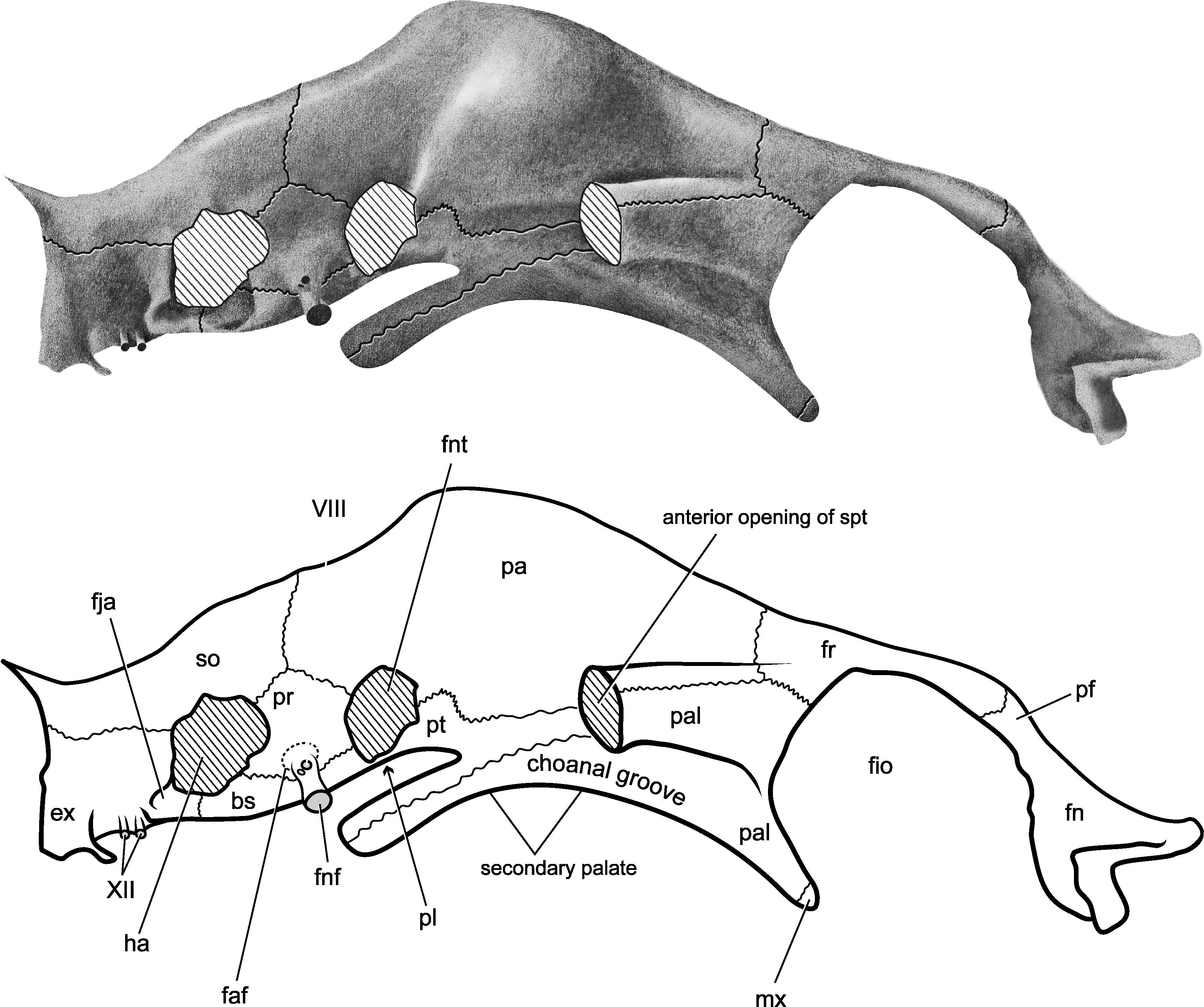
Fig. 1
Hamadachelys escuilliei Tong and Buffetaut, 1996. Partially restored skull based on MDE T03 and AMNH 30644. A, dorsal; B, ventral; C, lateral. [C. Facella, del.]
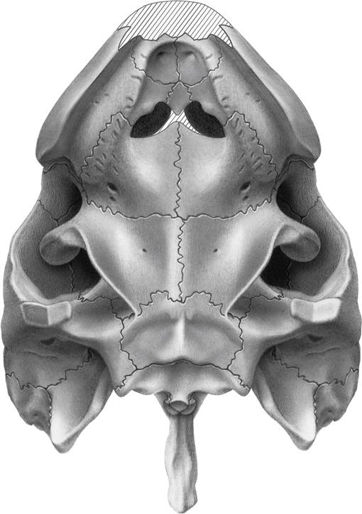
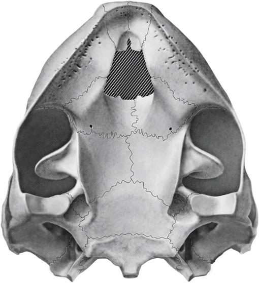
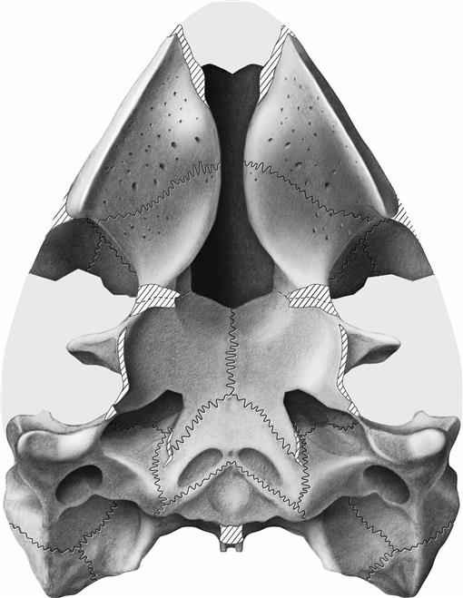
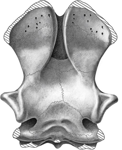
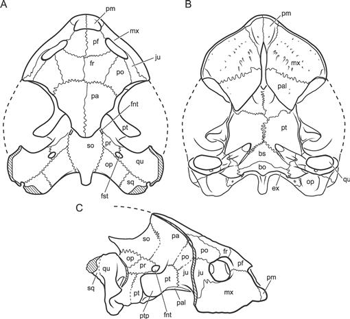
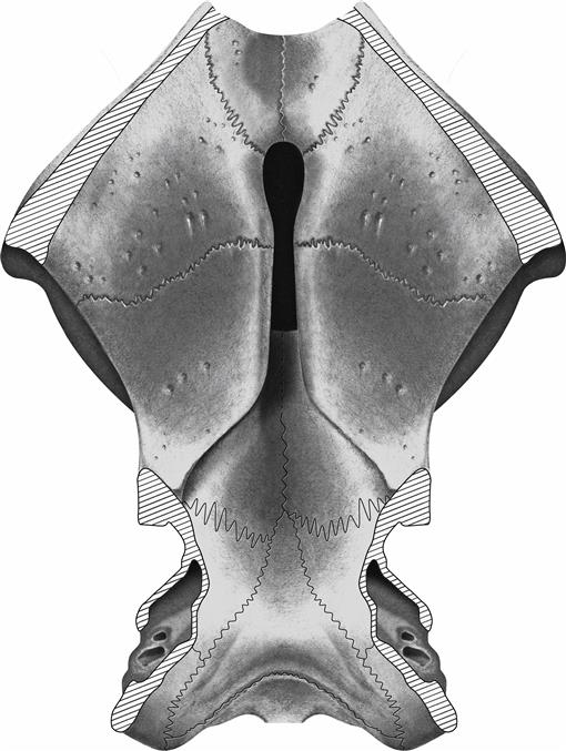
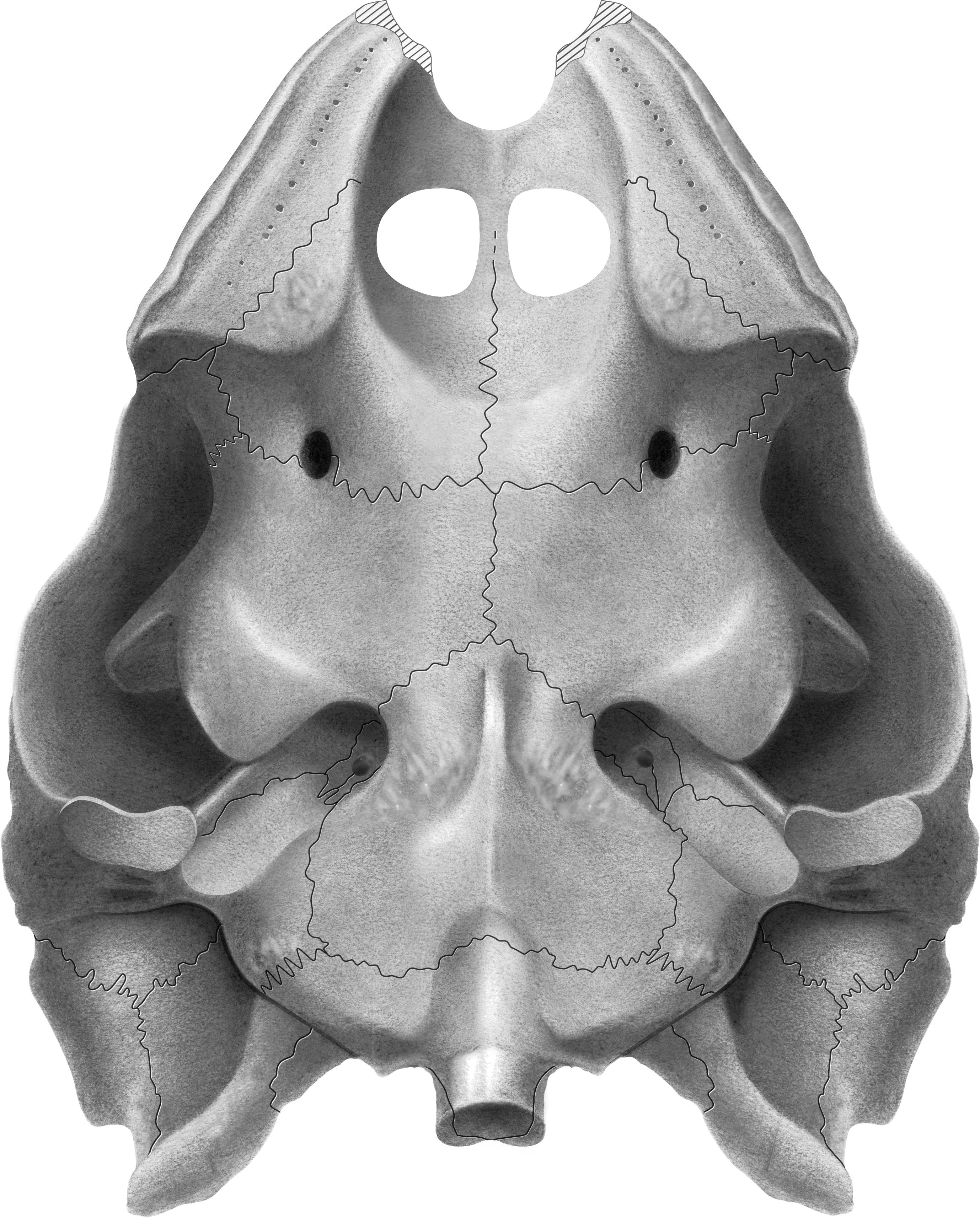
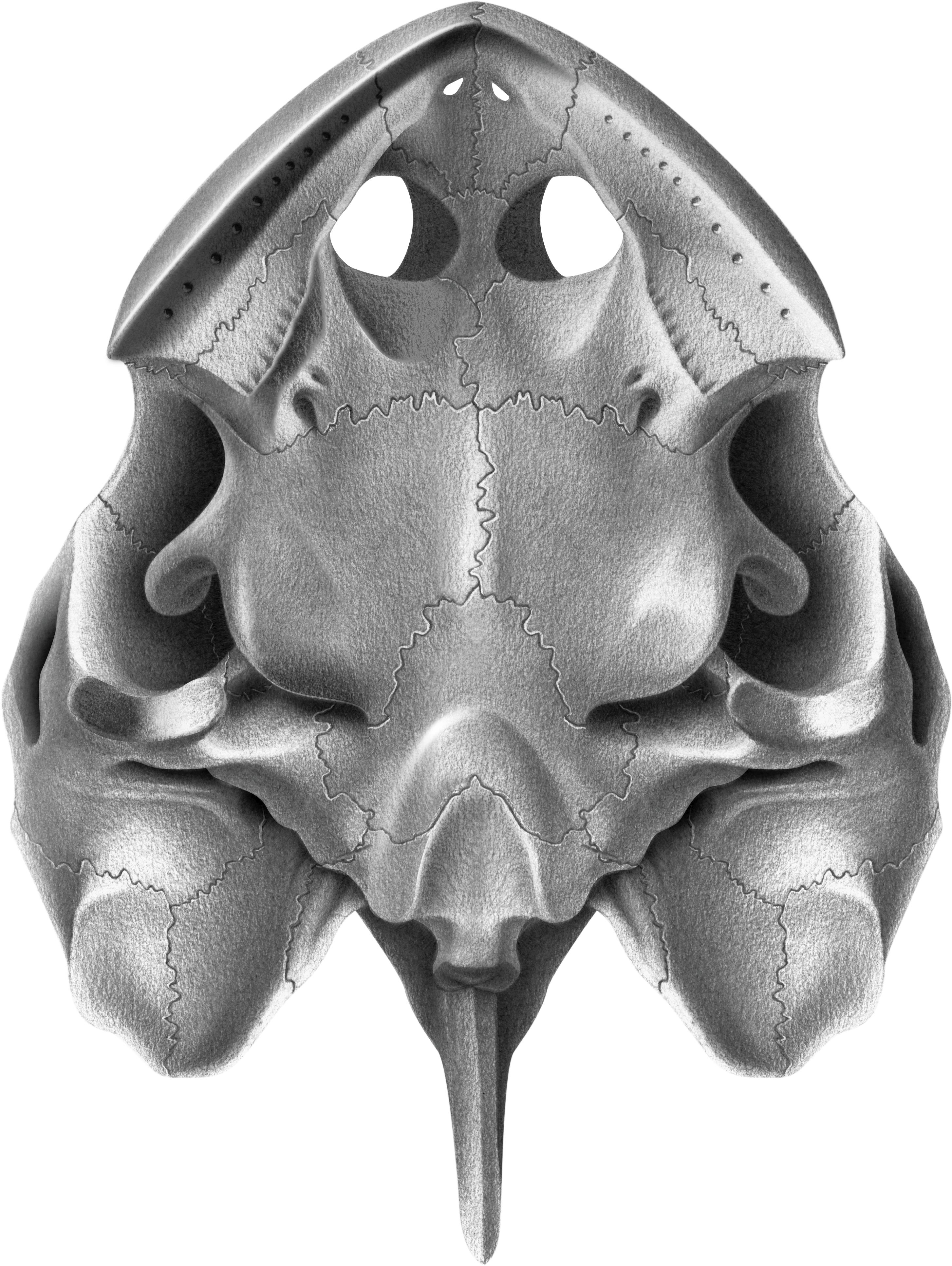
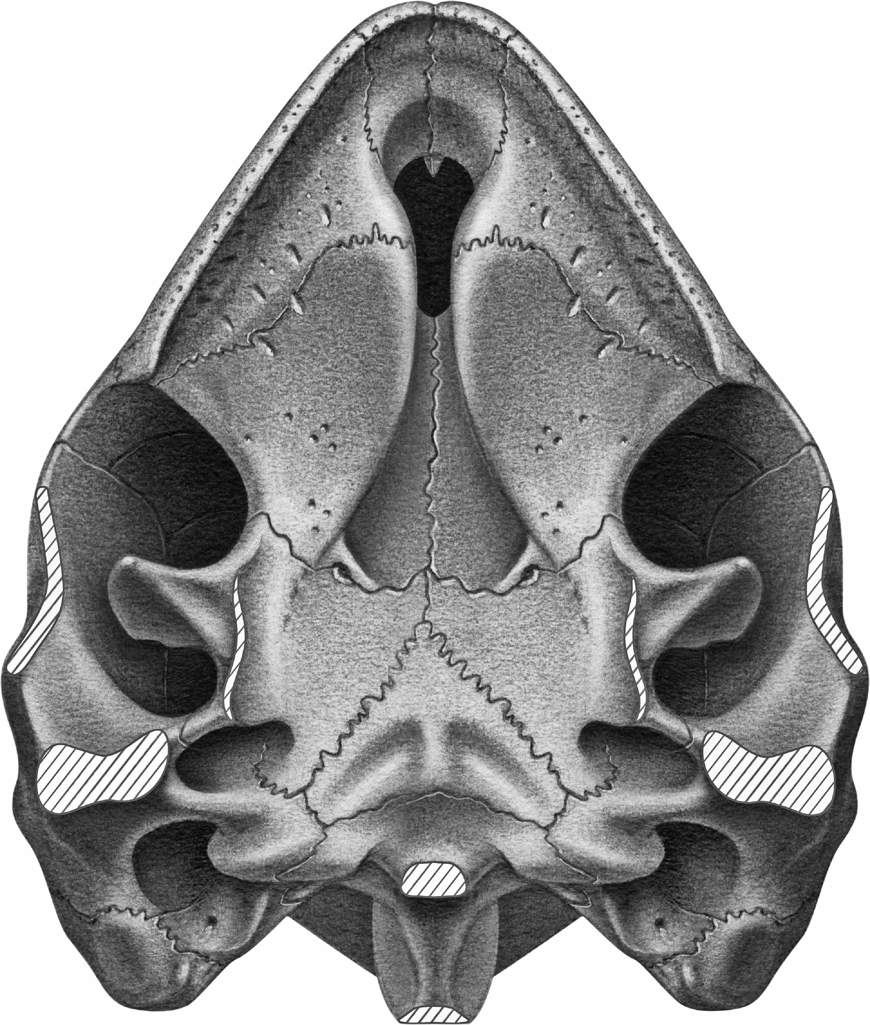
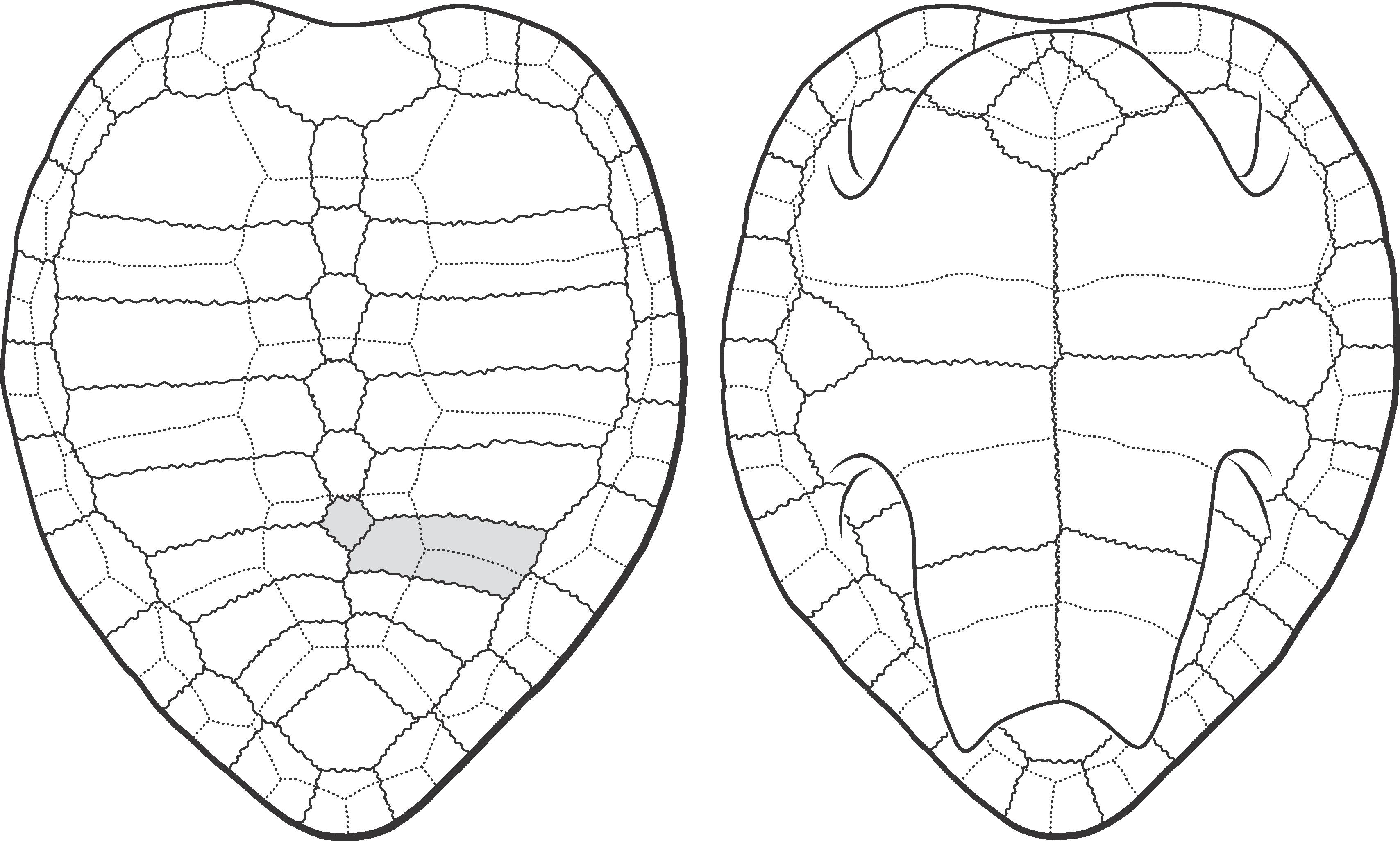
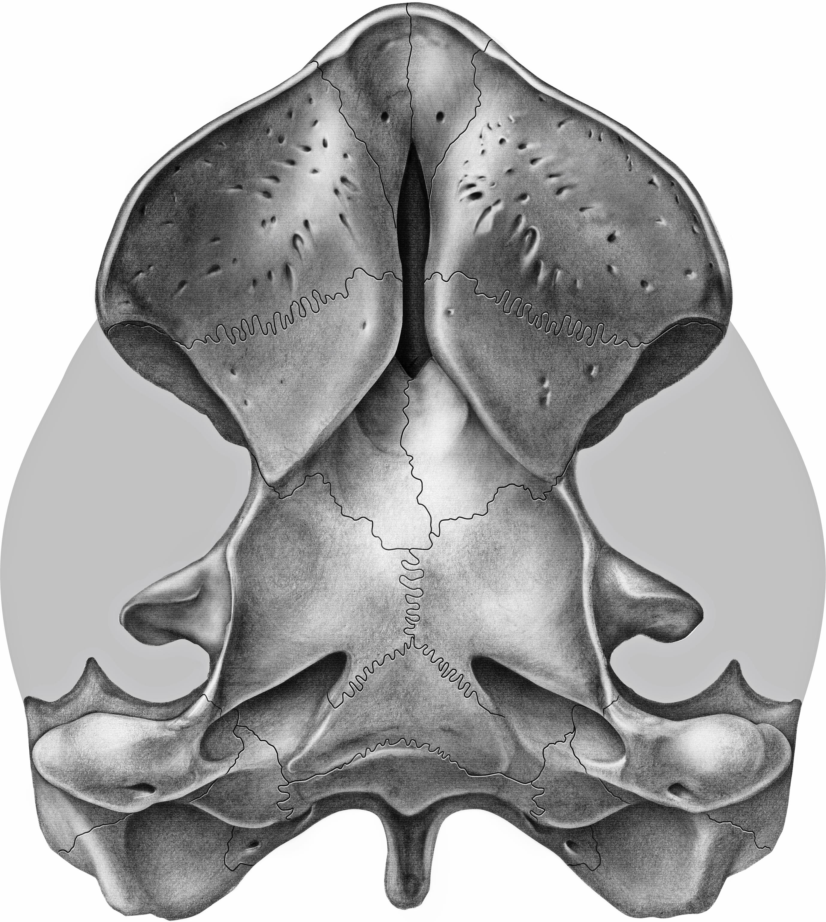
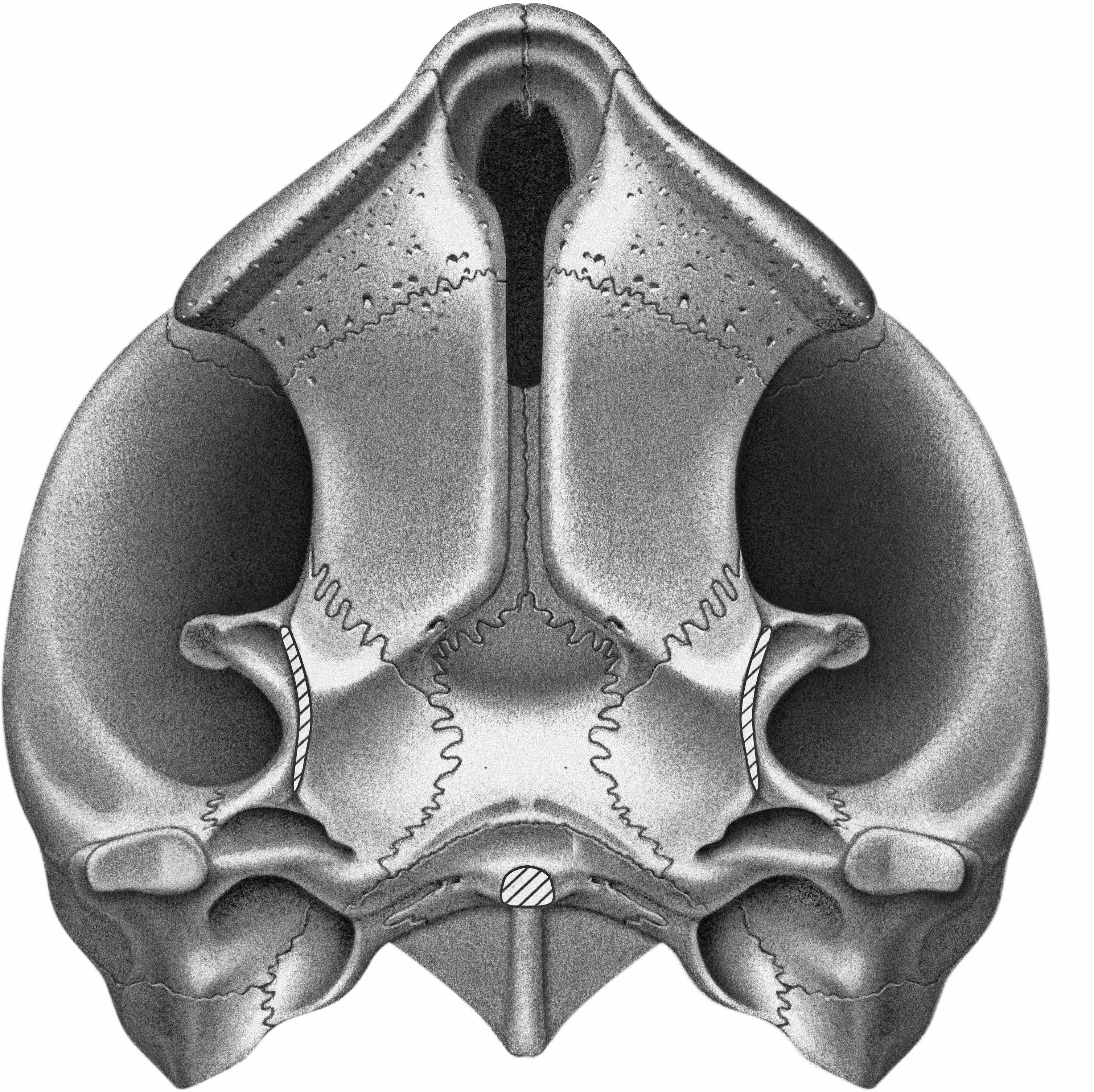
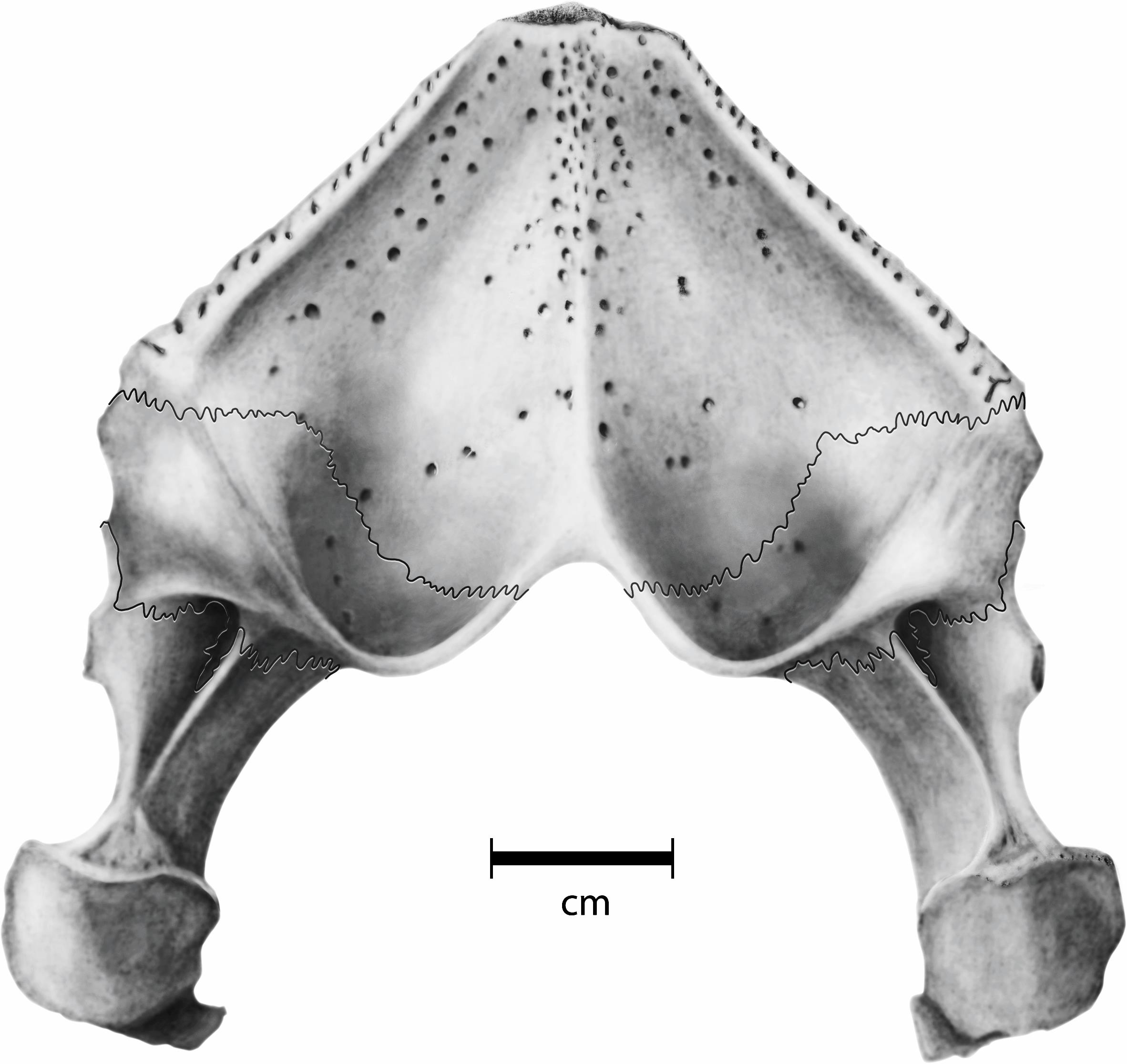
Fig. 2
Hamadachelys escuilliei Tong and Buffetaut, 1996. Partially restored ventral view based on MDE T03 and AMNH 30644. [A. Phillips, del.]
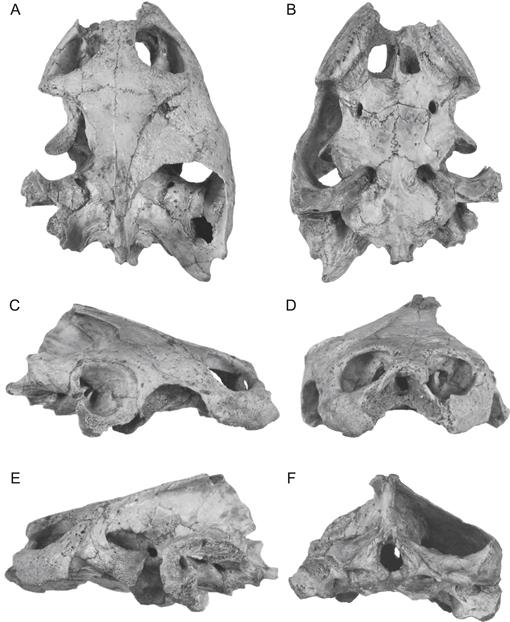
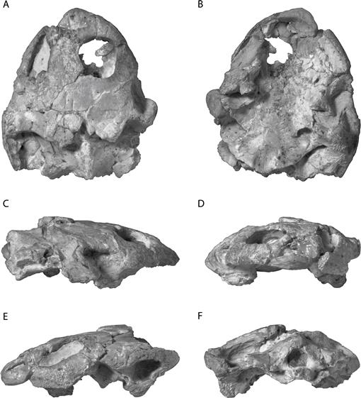
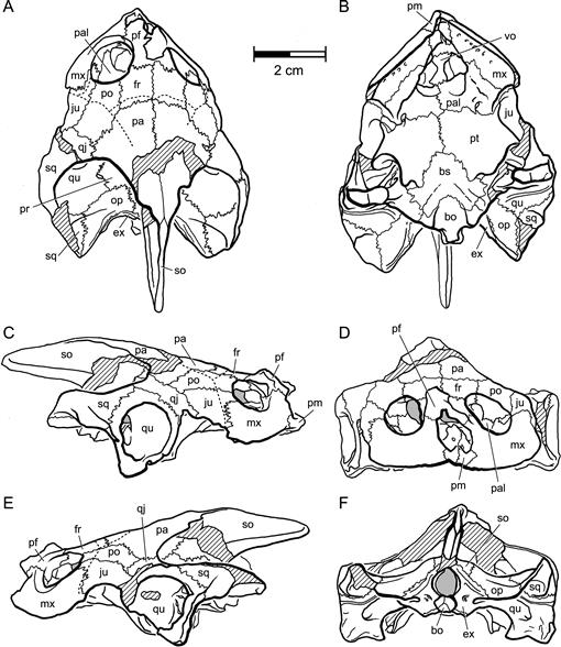
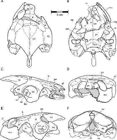
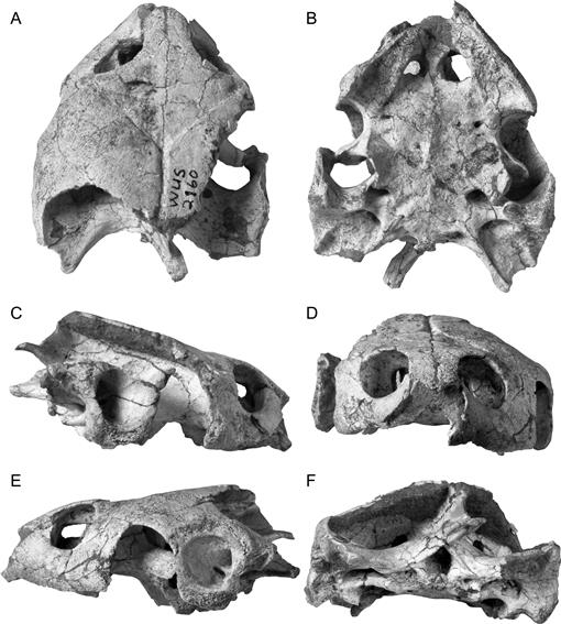
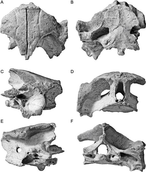
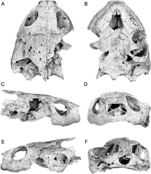
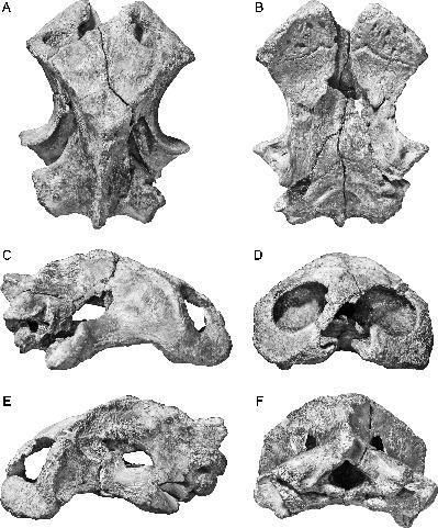
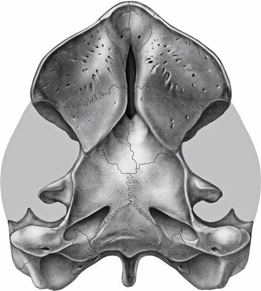
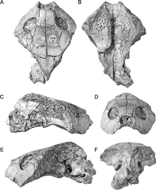
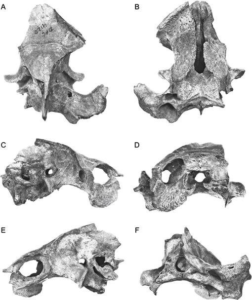
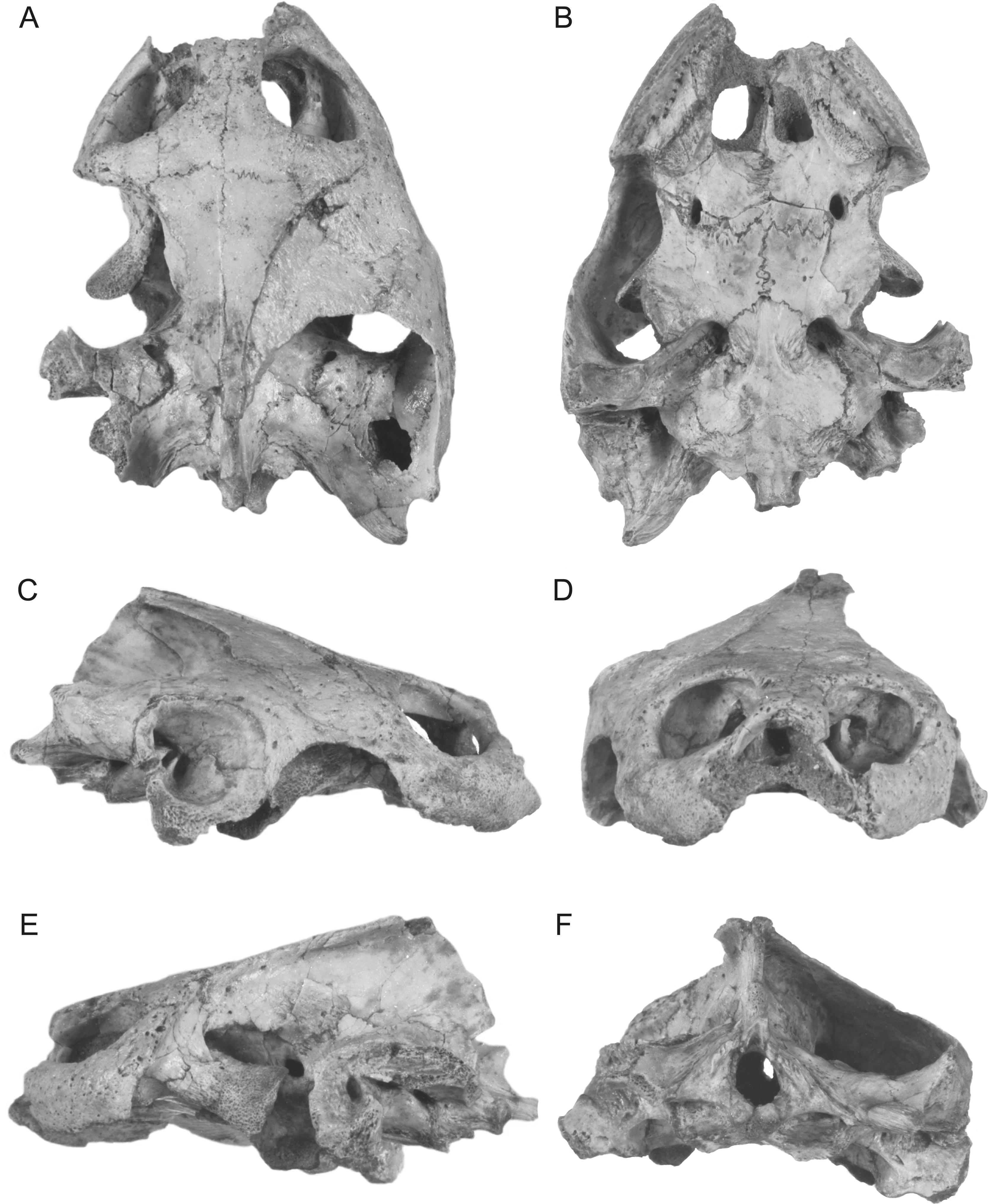
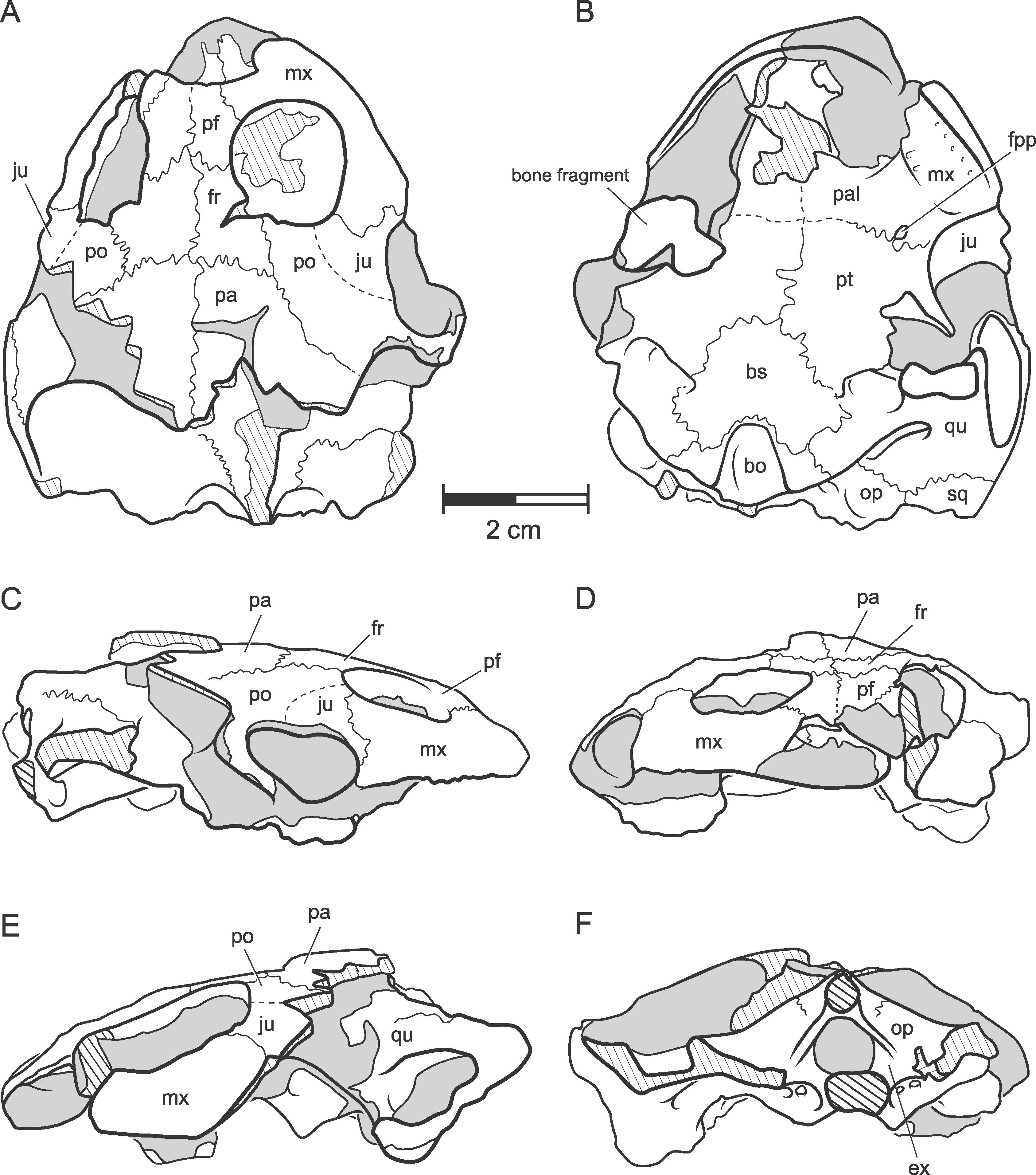
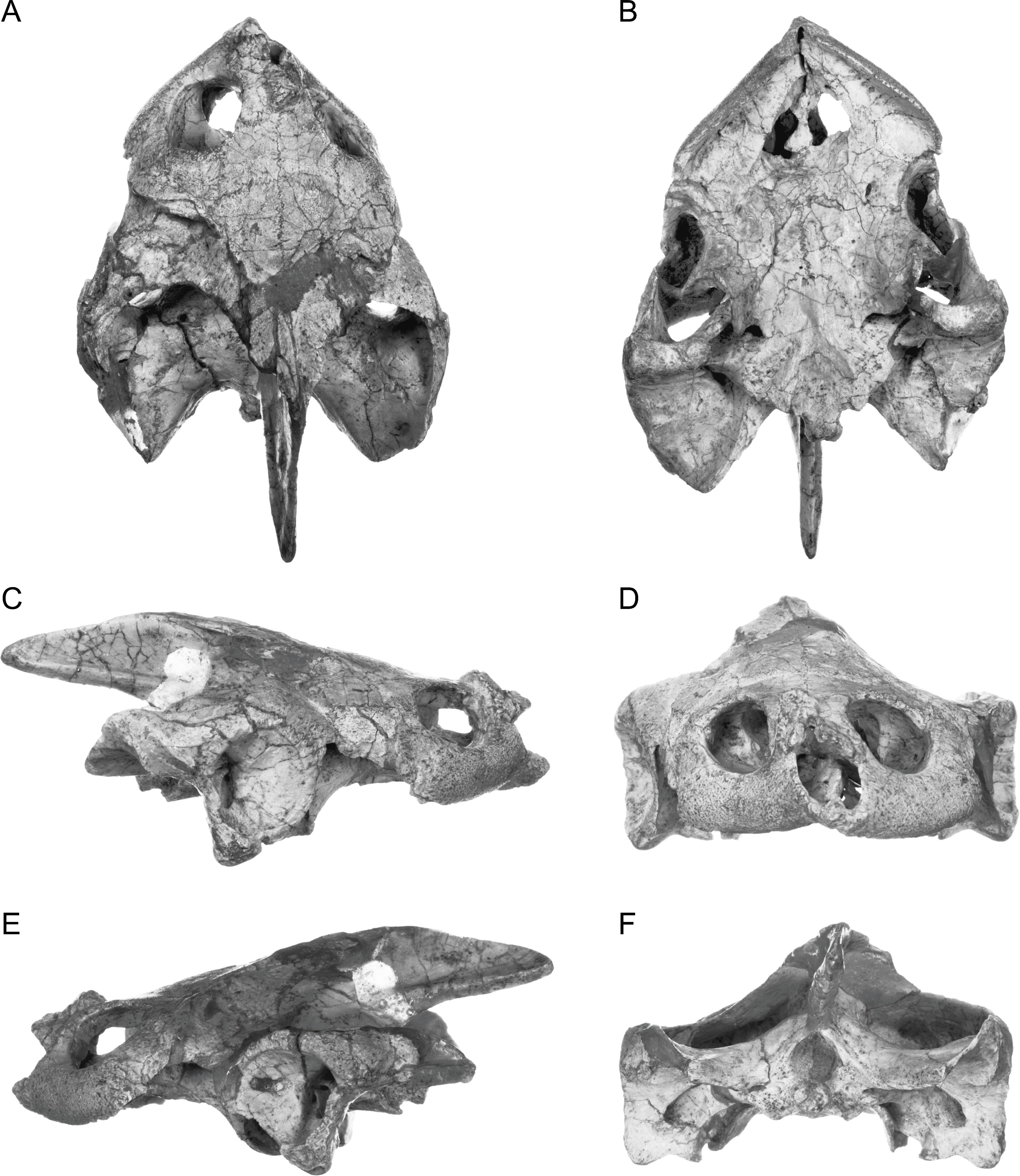
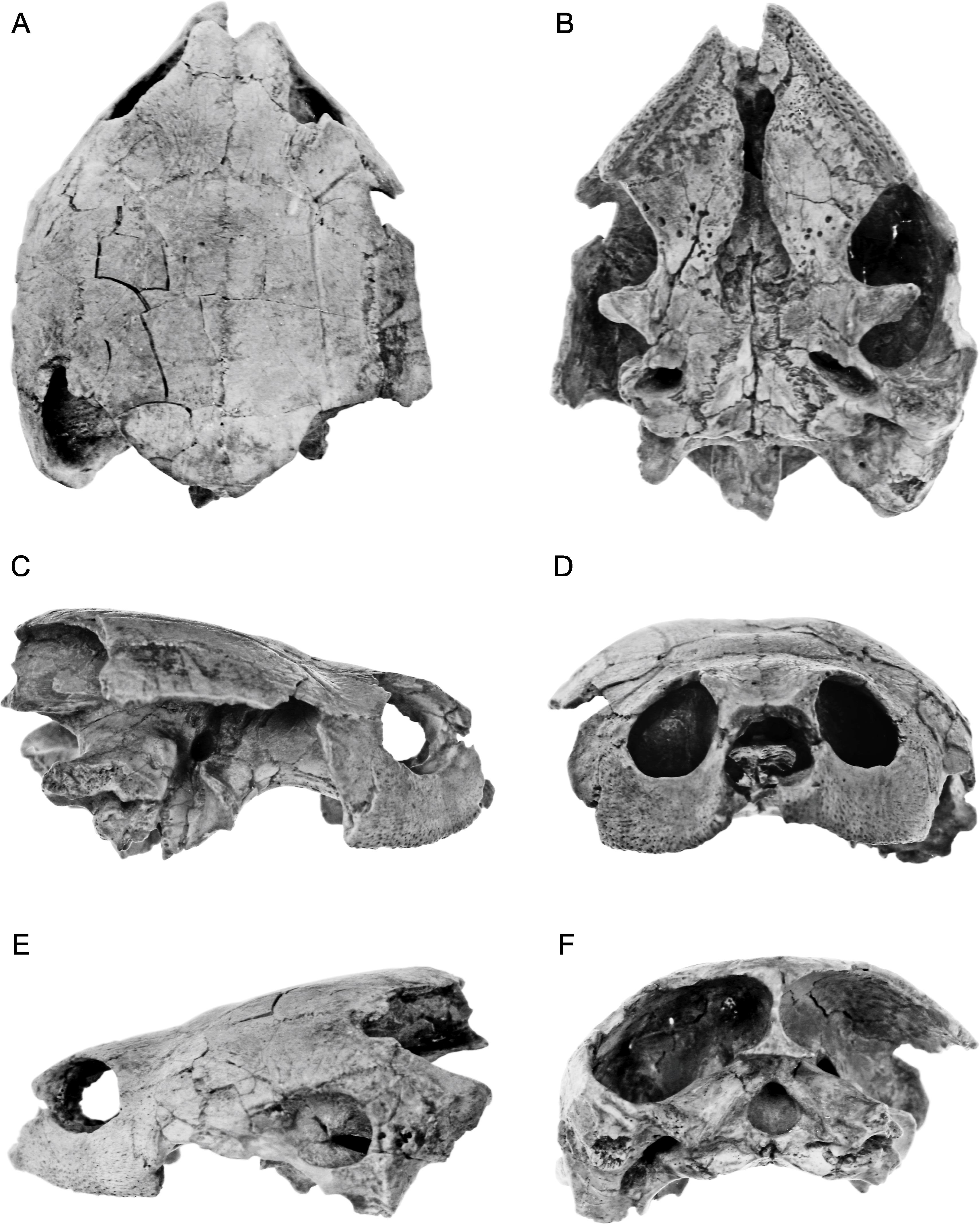
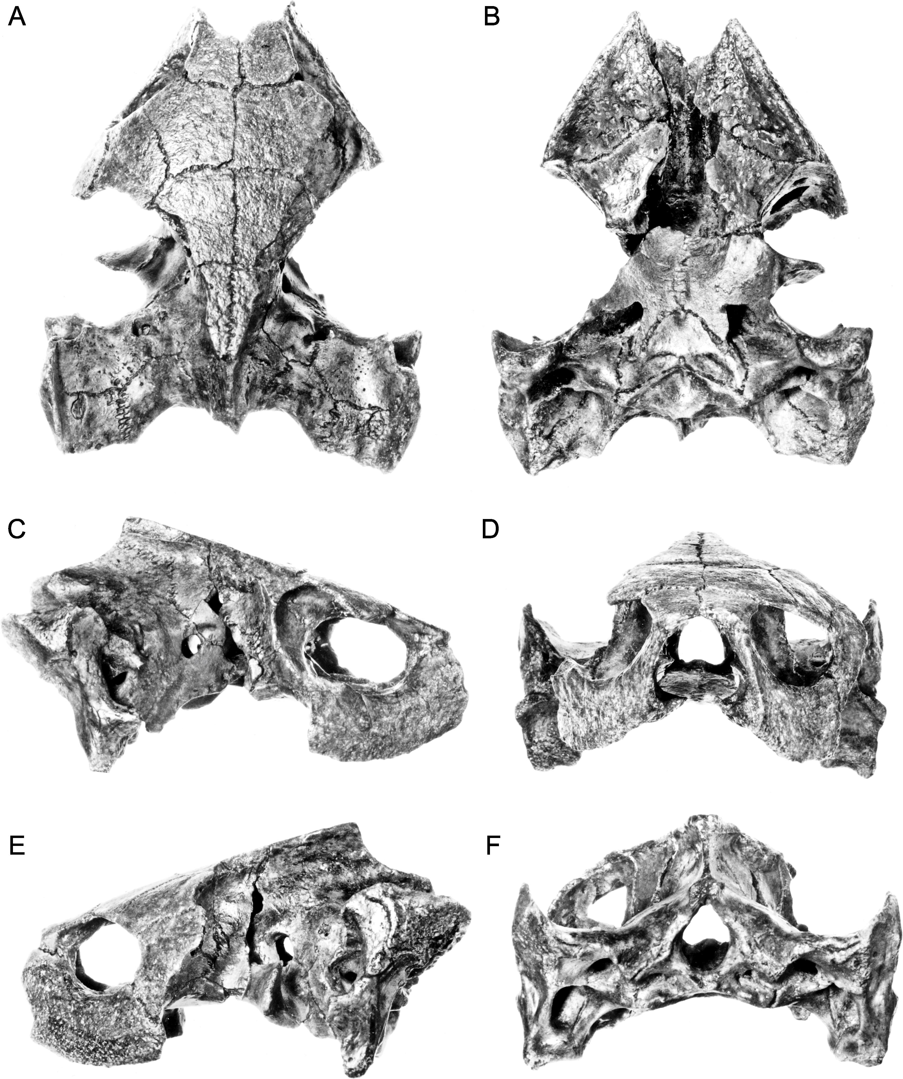
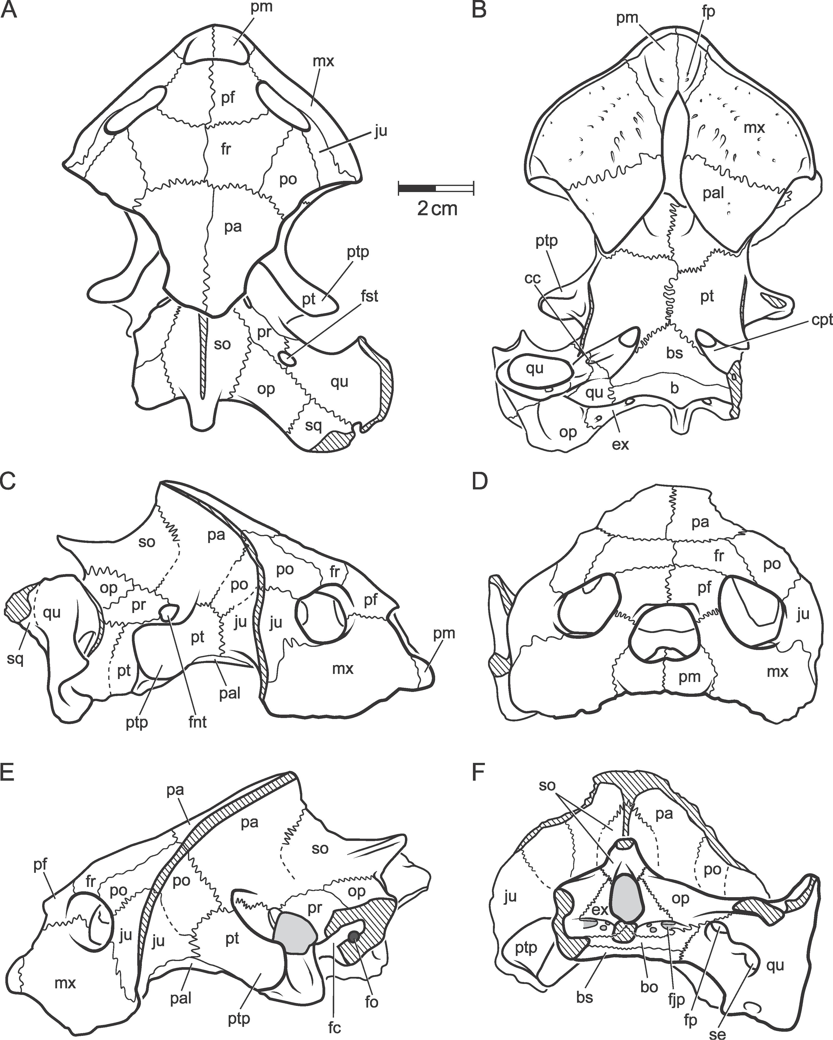
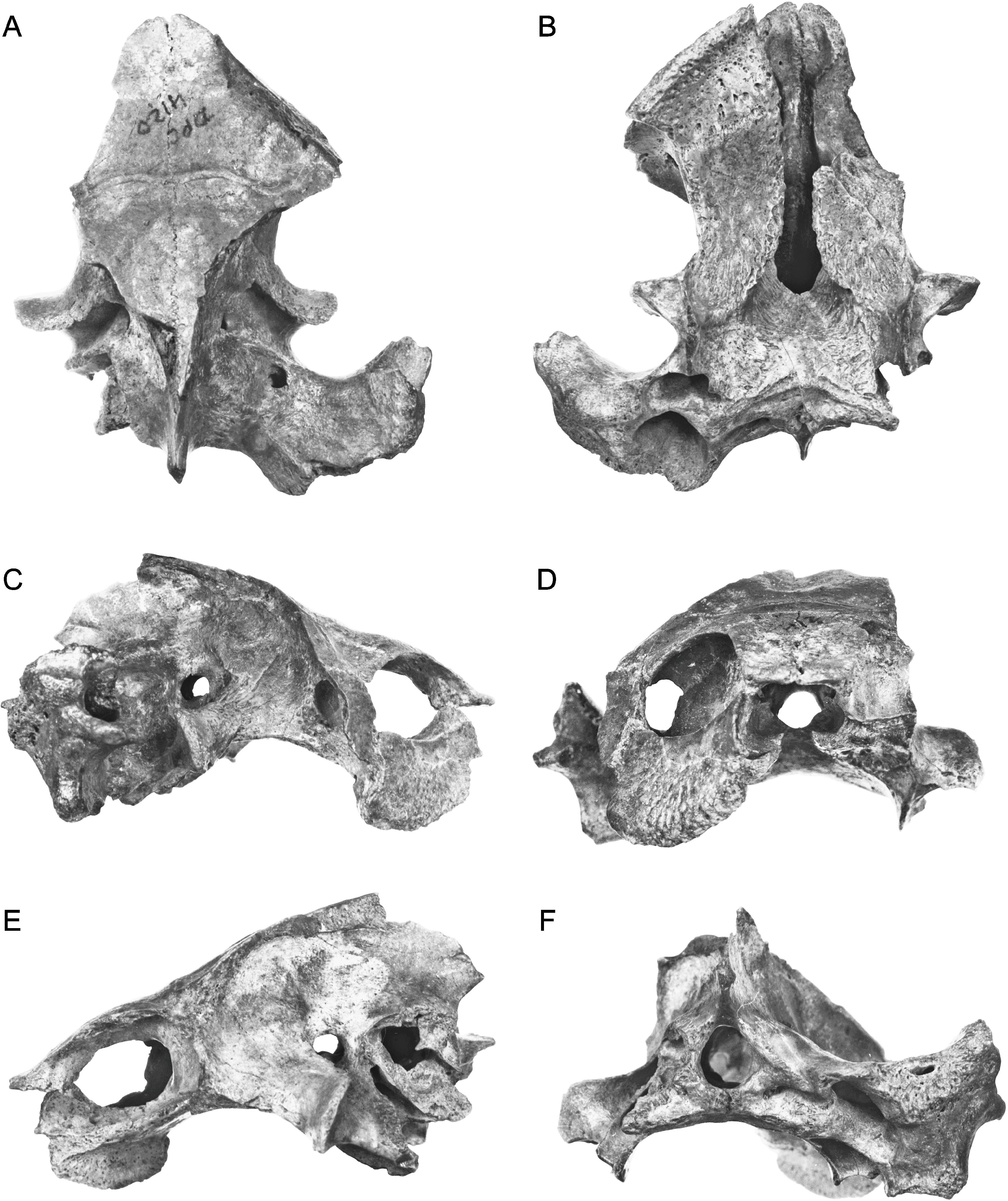
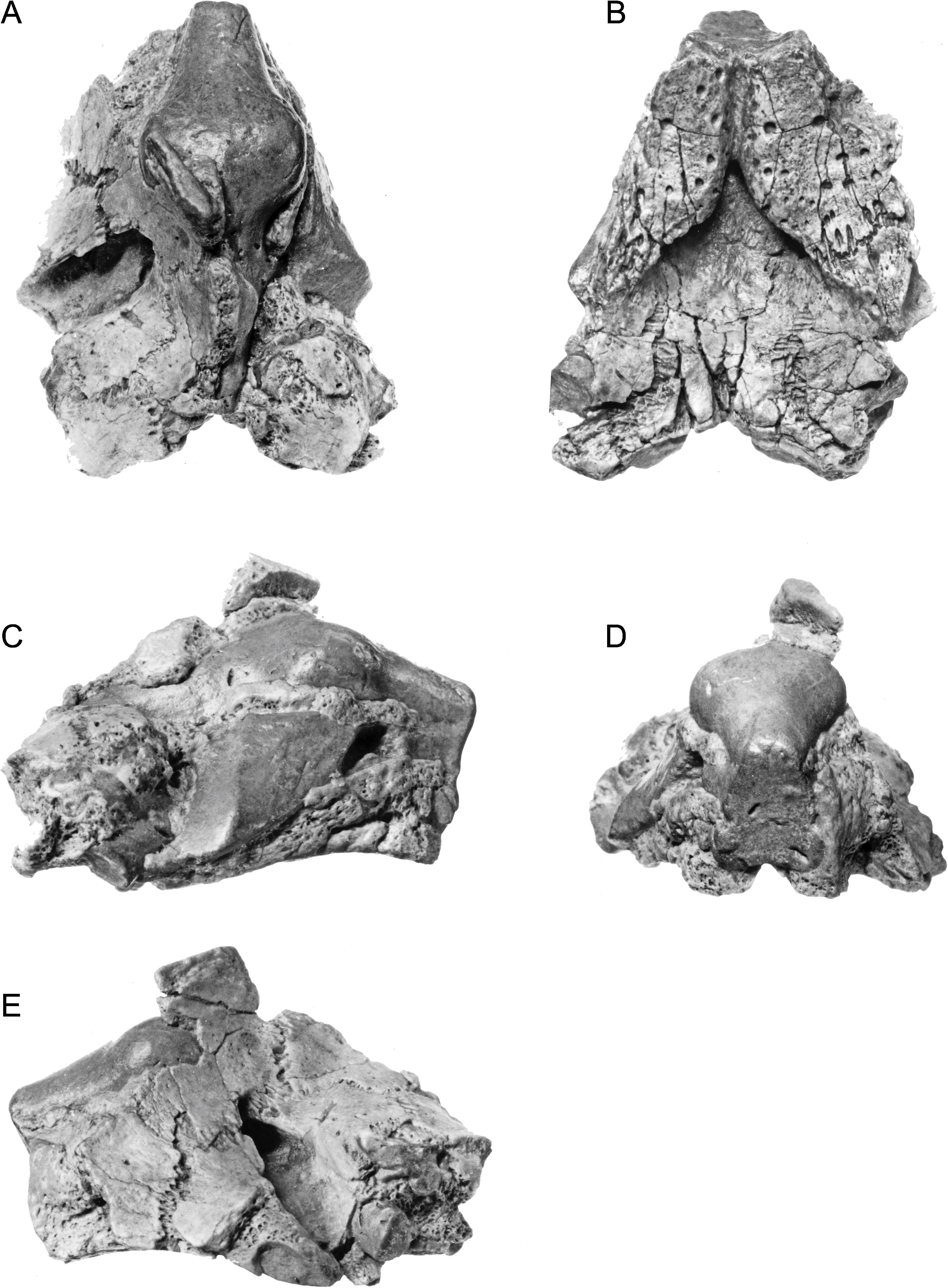
Fig. 3
Hamadachelys escuilliei Tong and Buffetaut, 1996. MDE T03. A, dorsal; B, ventral; C, right lateral; D, anterior; E, left lateral; F, posterior. [A. Phillips, del.]
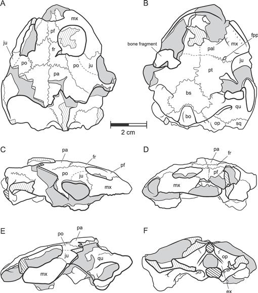
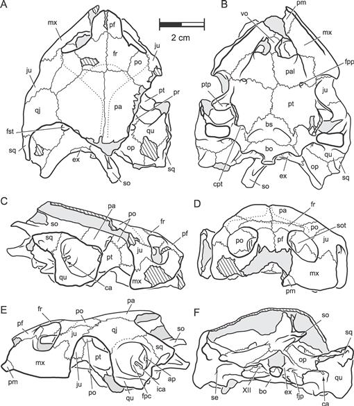
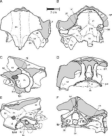
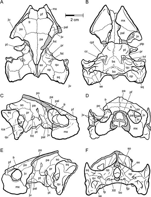
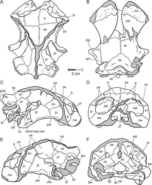
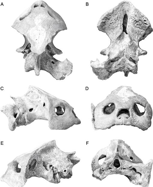
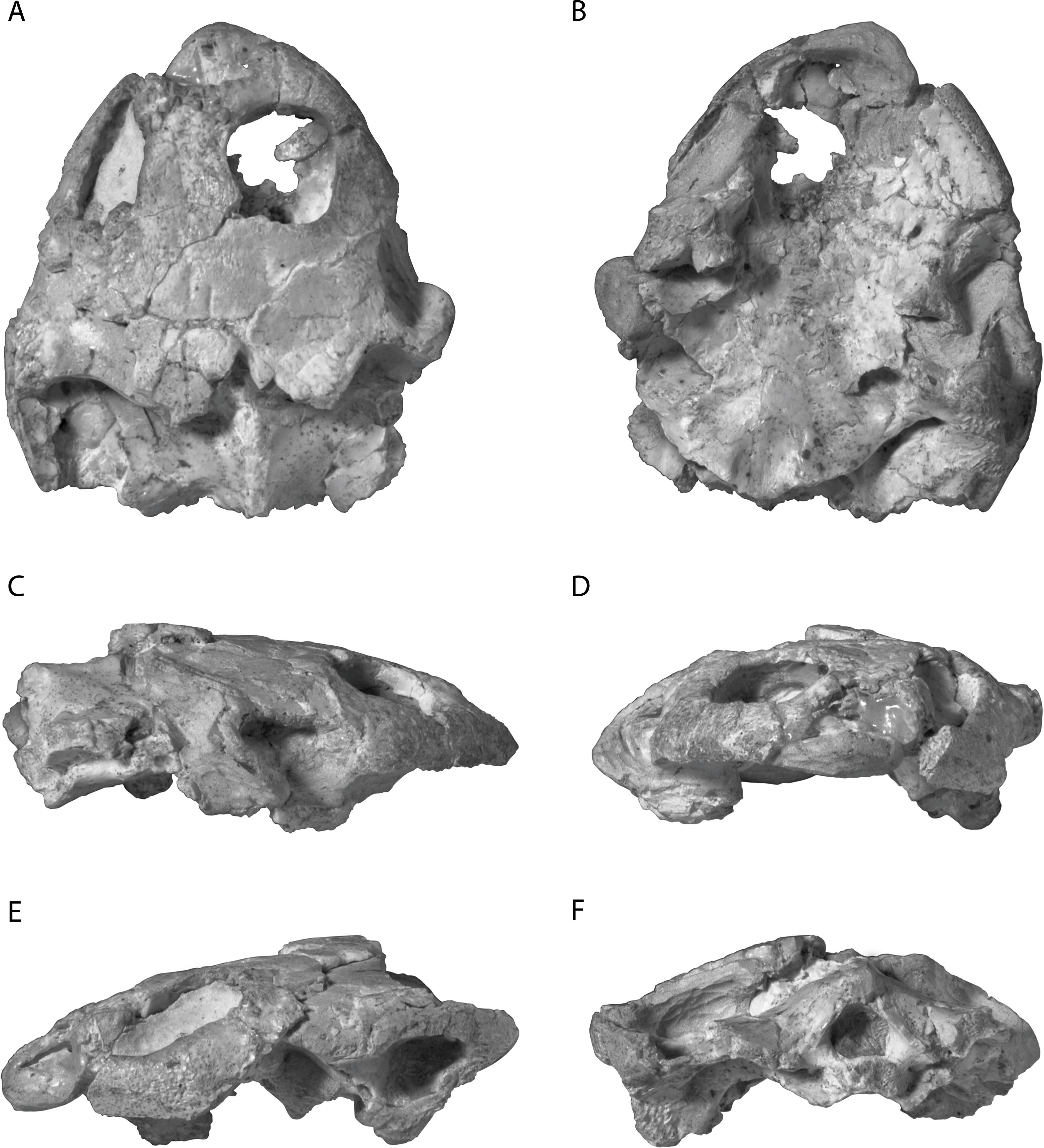
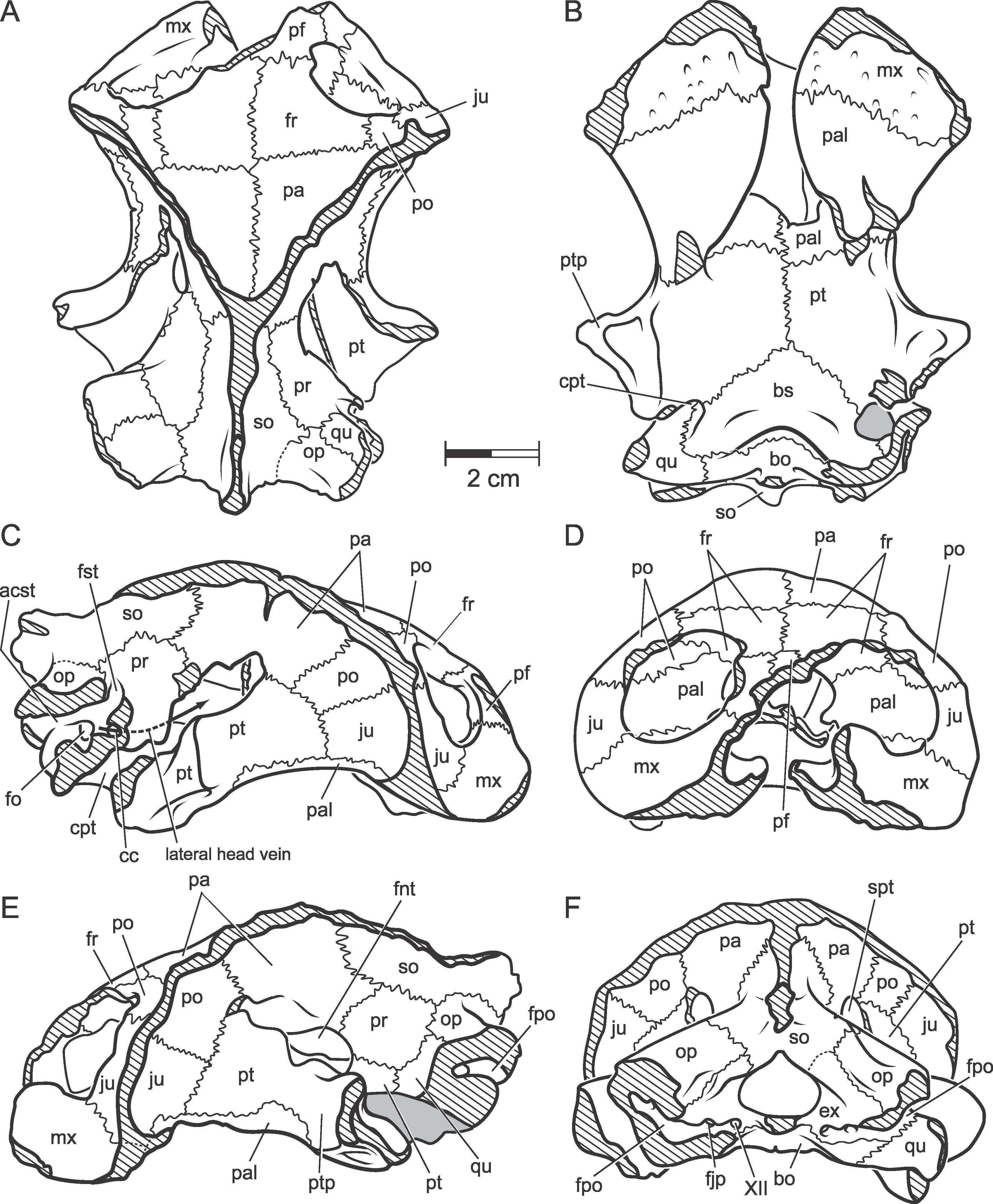
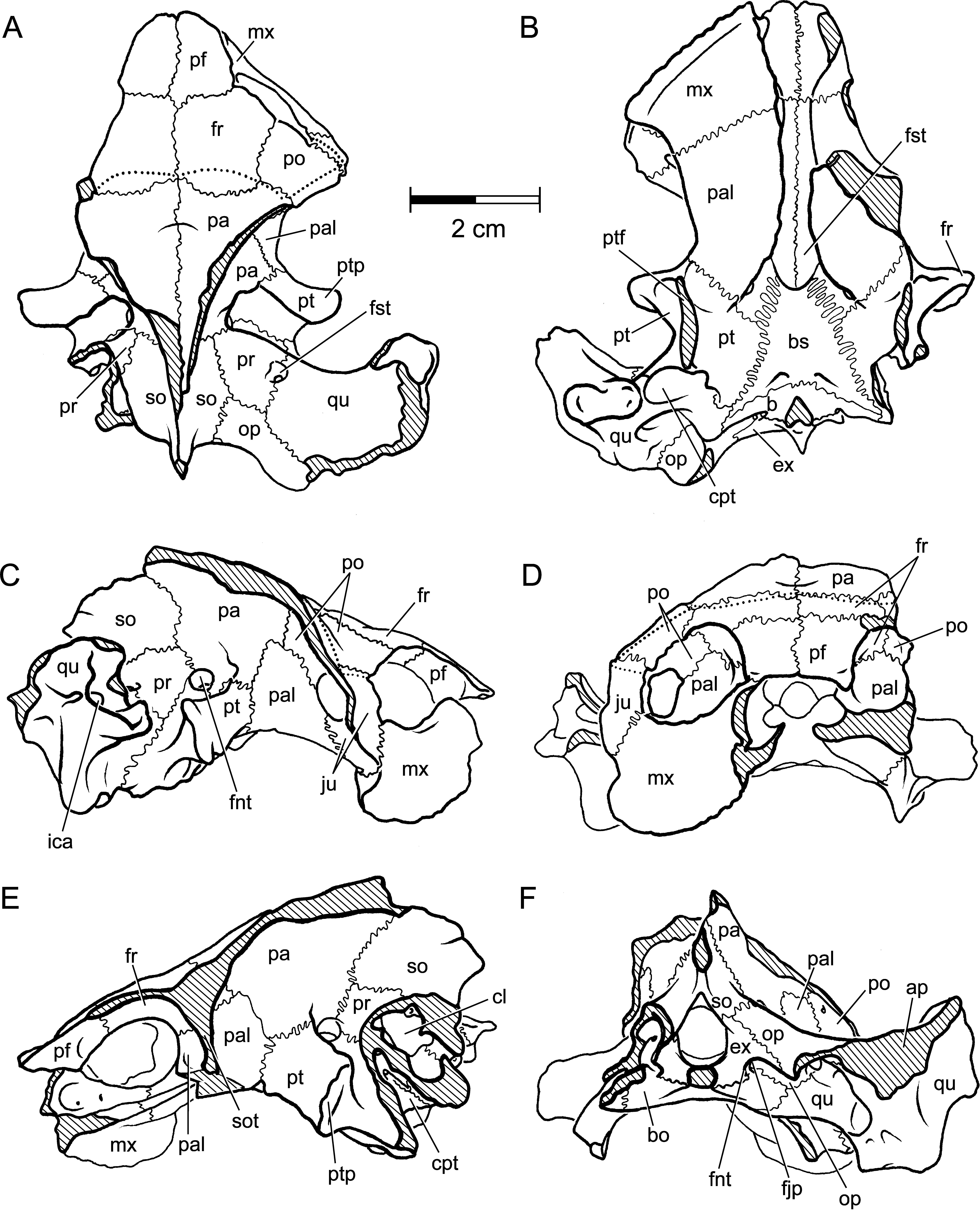
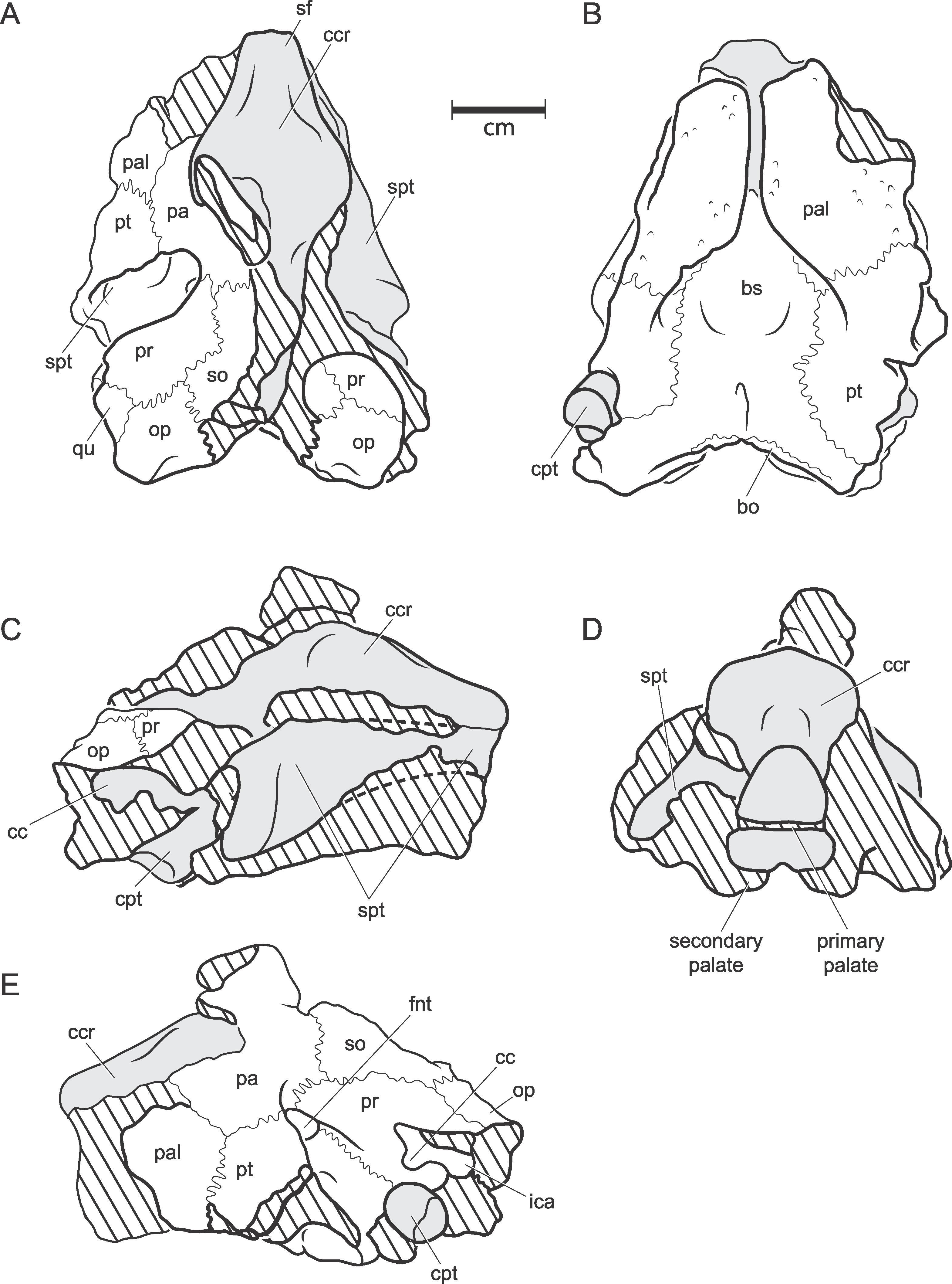
Fig. 4
Hamadachelys escuilliei Tong and Buffetaut, 1996. MDE T03. A, dorsal; B, ventral; C, right lateral; D, anterior; E, left lateral; F, posterior. [A. Phillips, del.]
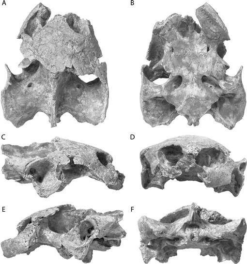
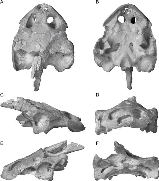
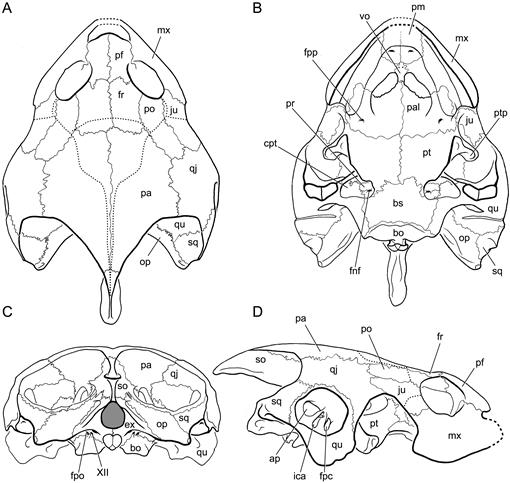
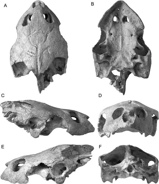
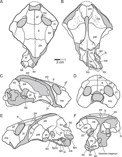
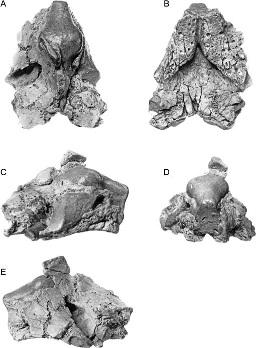
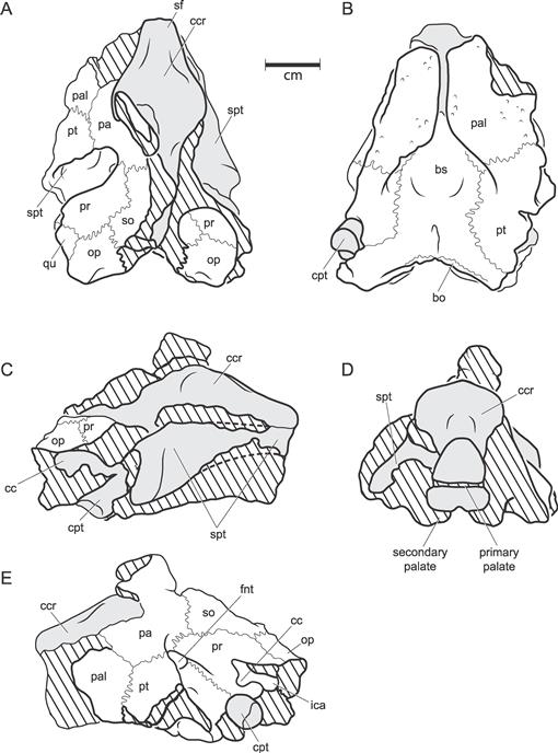
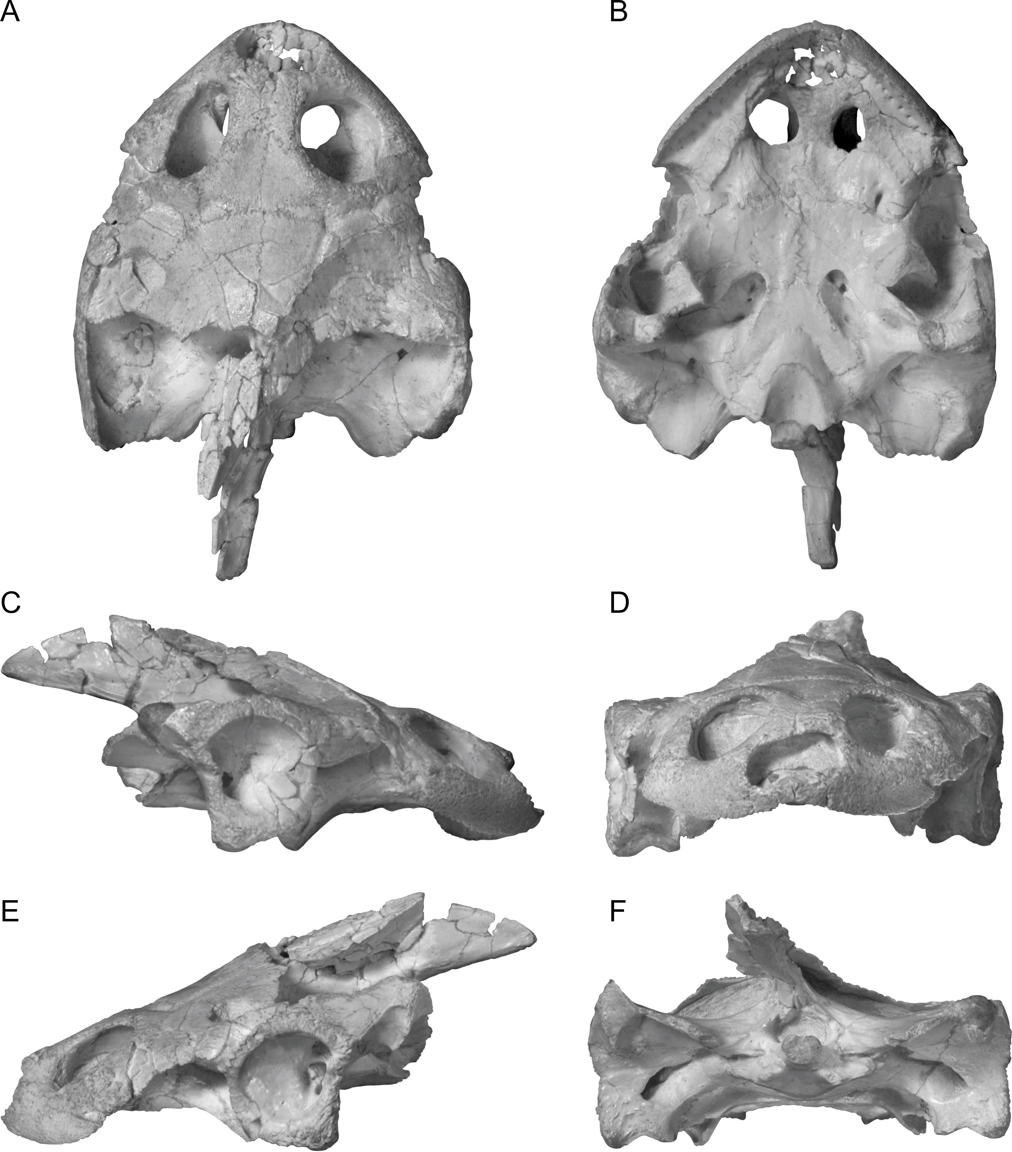
Fig. 5
Hamadachelys escuilliei Tong and Buffetaut, 1996. AMNH 30644. A, dorsal; B, ventral; C, right lateral; D, anterior; E, left lateral; F, posterior. [C. Facella del.]
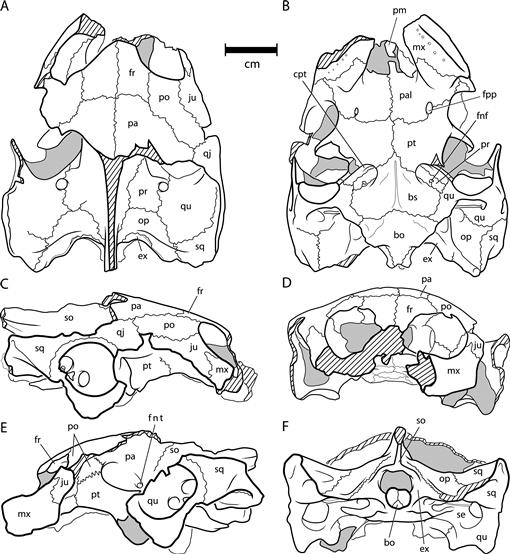
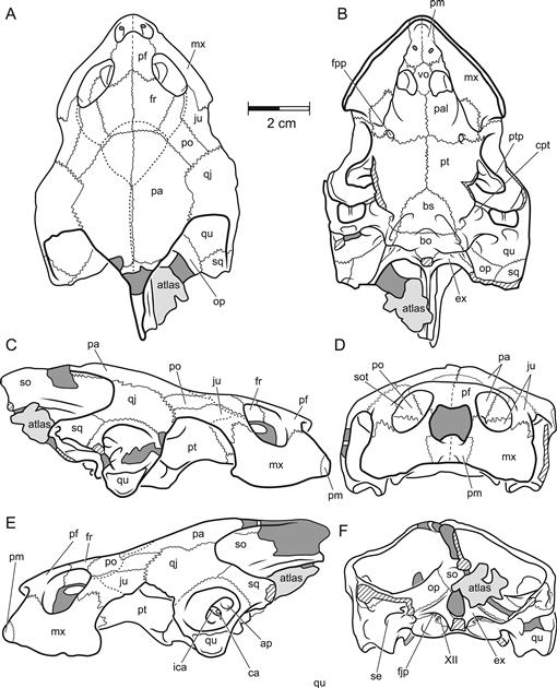
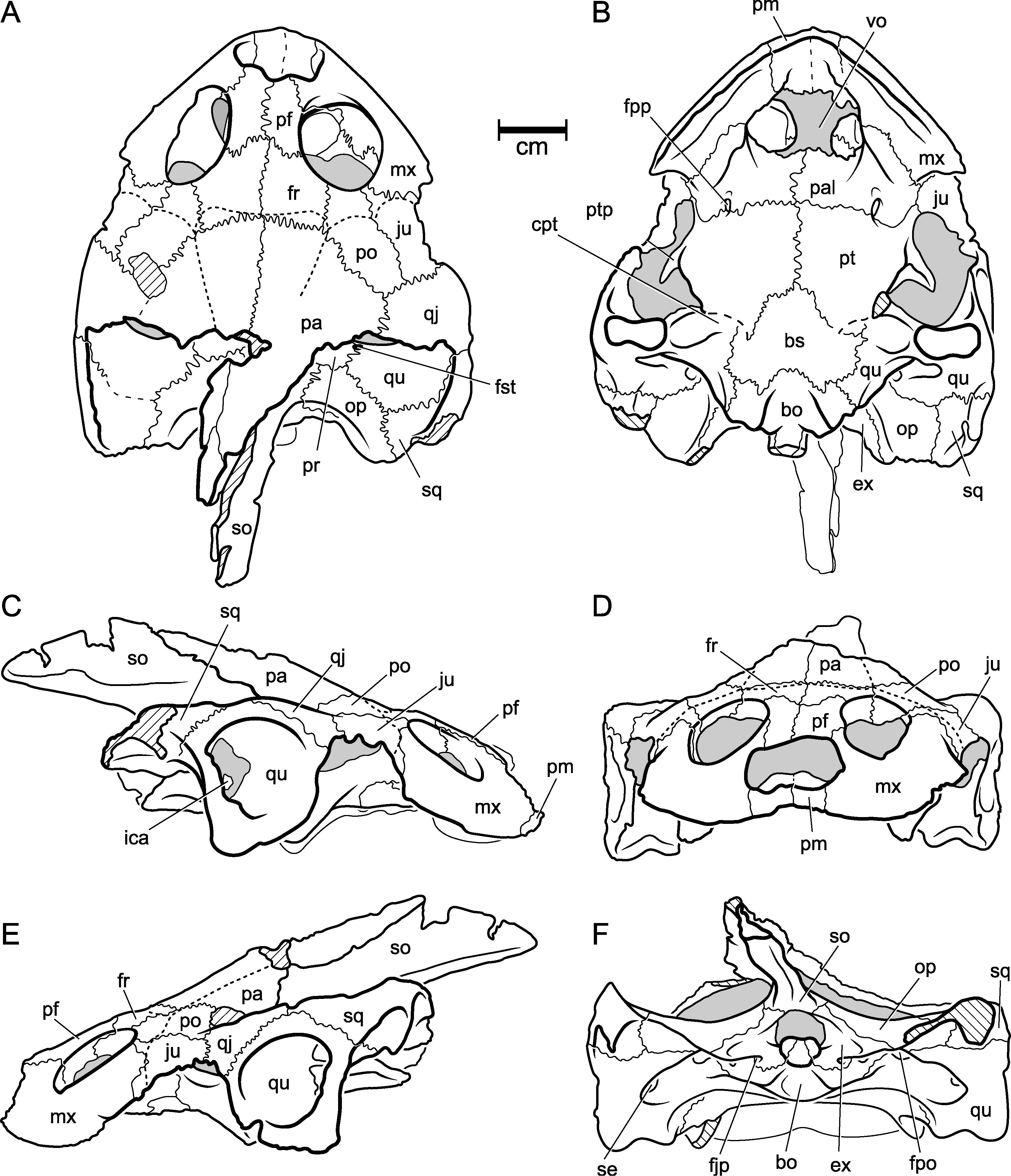
Fig. 6
Hamadachelys escuilliei Tong and Buffetaut, 1996. AMNH 30644. A, dorsal; B, ventral; C, right lateral; D, anterior; E, left lateral; F, posterior. [C. Facella del.]
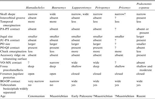
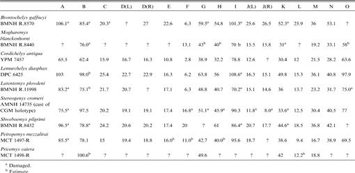
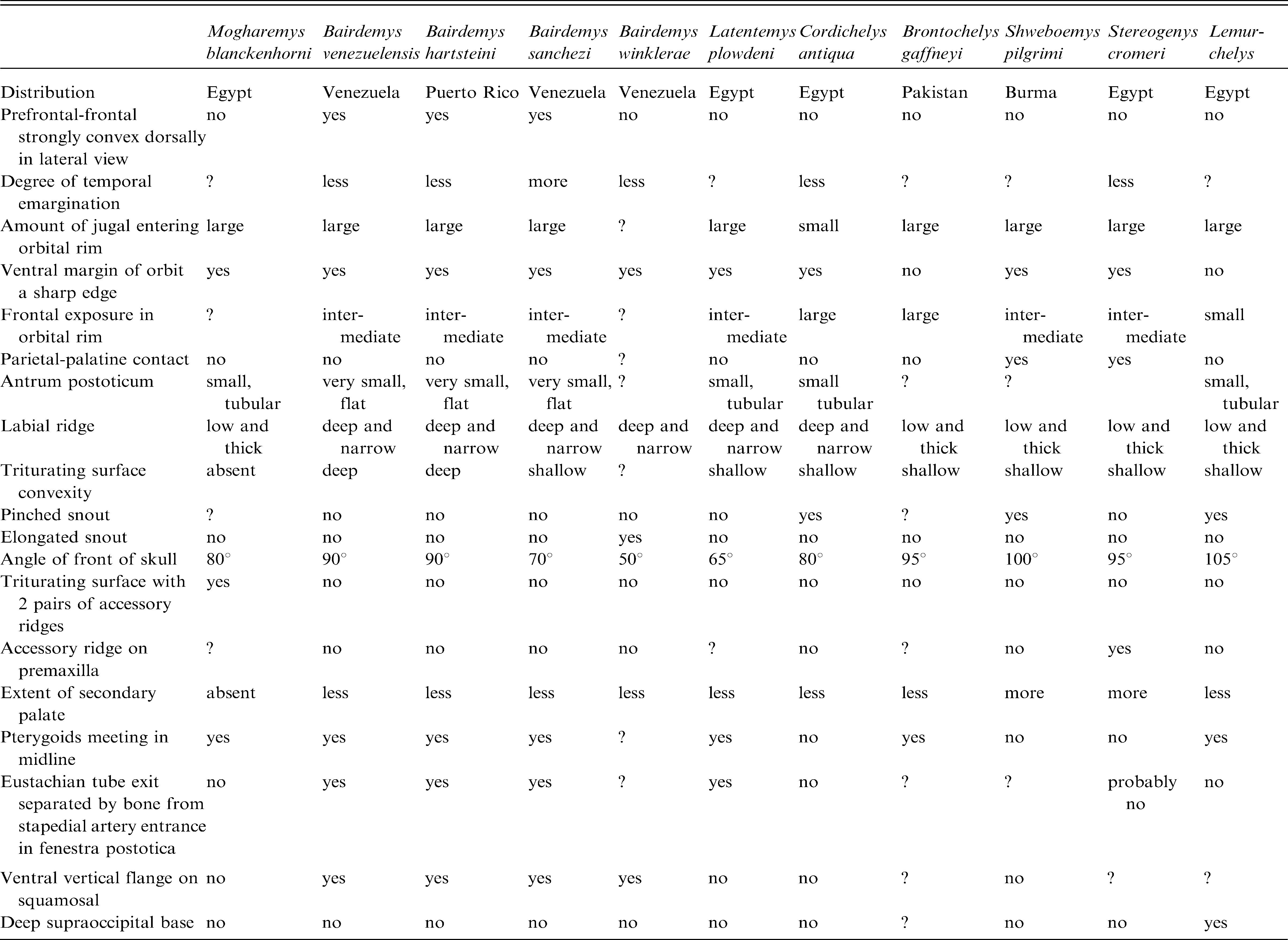
TABLE 1
Comparison of Skulls of Mesozoic and Early Tertiary Podocnemididae
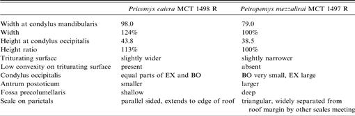
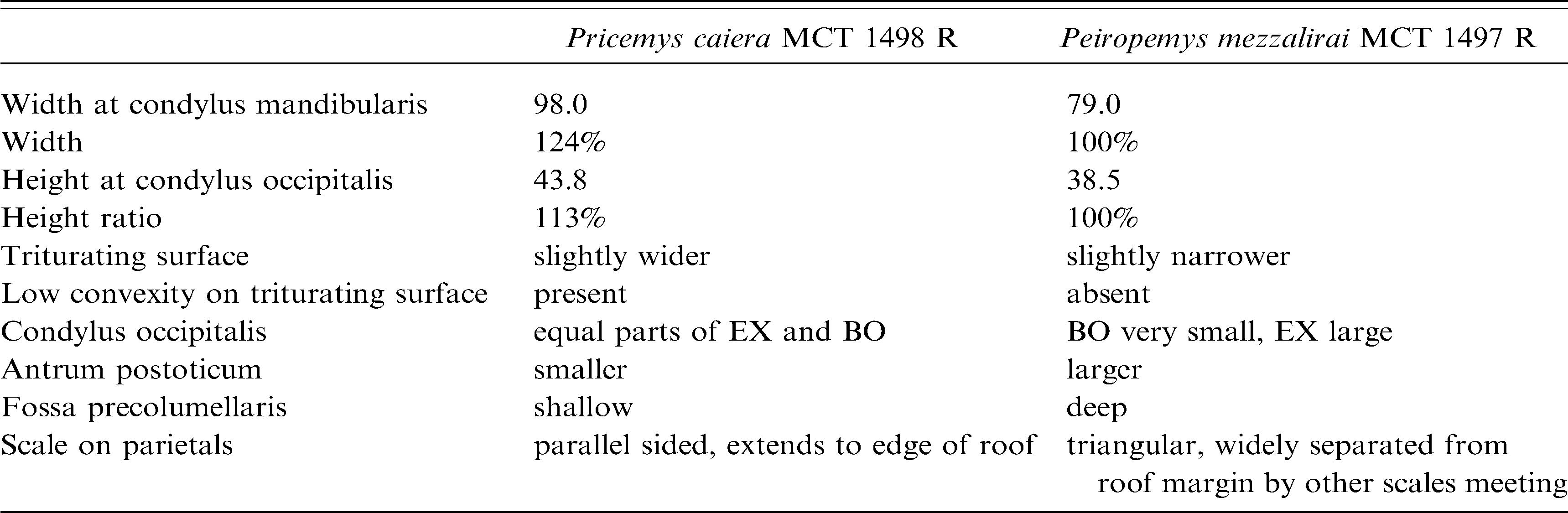
TABLE 2
Comparison of Peirópolis Skulls (measurements in mm)
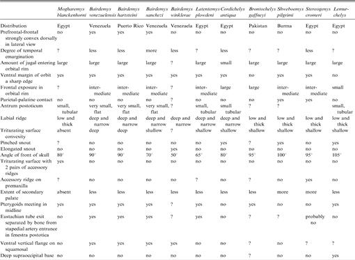
TABLE 3
Species of the Tribe Stereogenyini
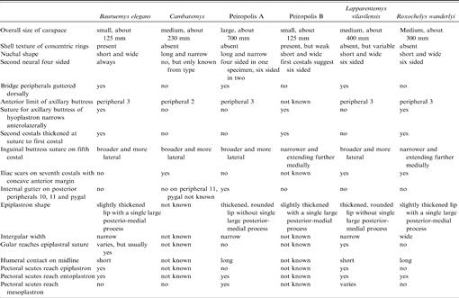
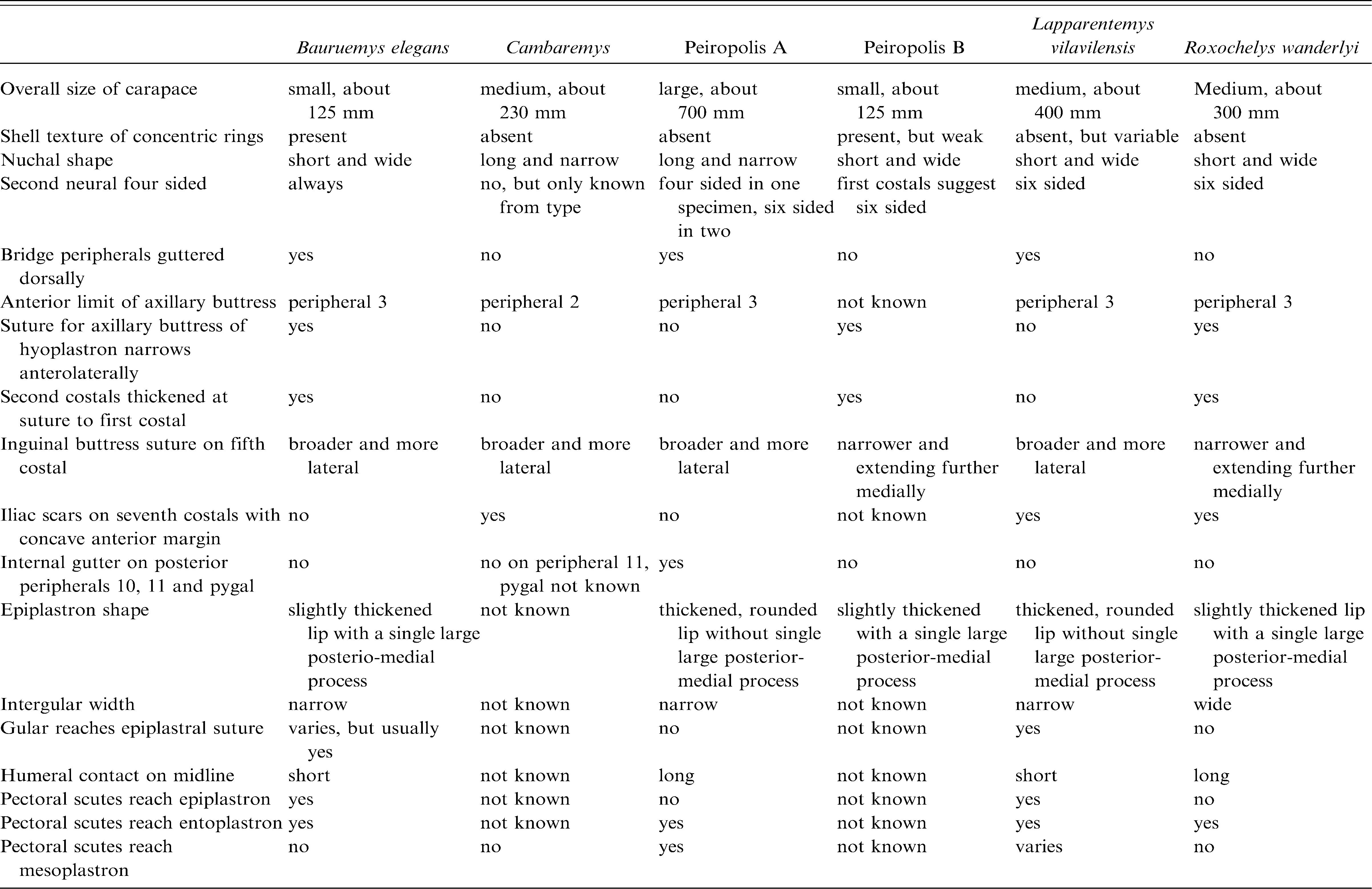
TABLE 4
Comparison of Shells of Six South American Late Cretaceous and Early Tertiary Podocnemidid Turtles
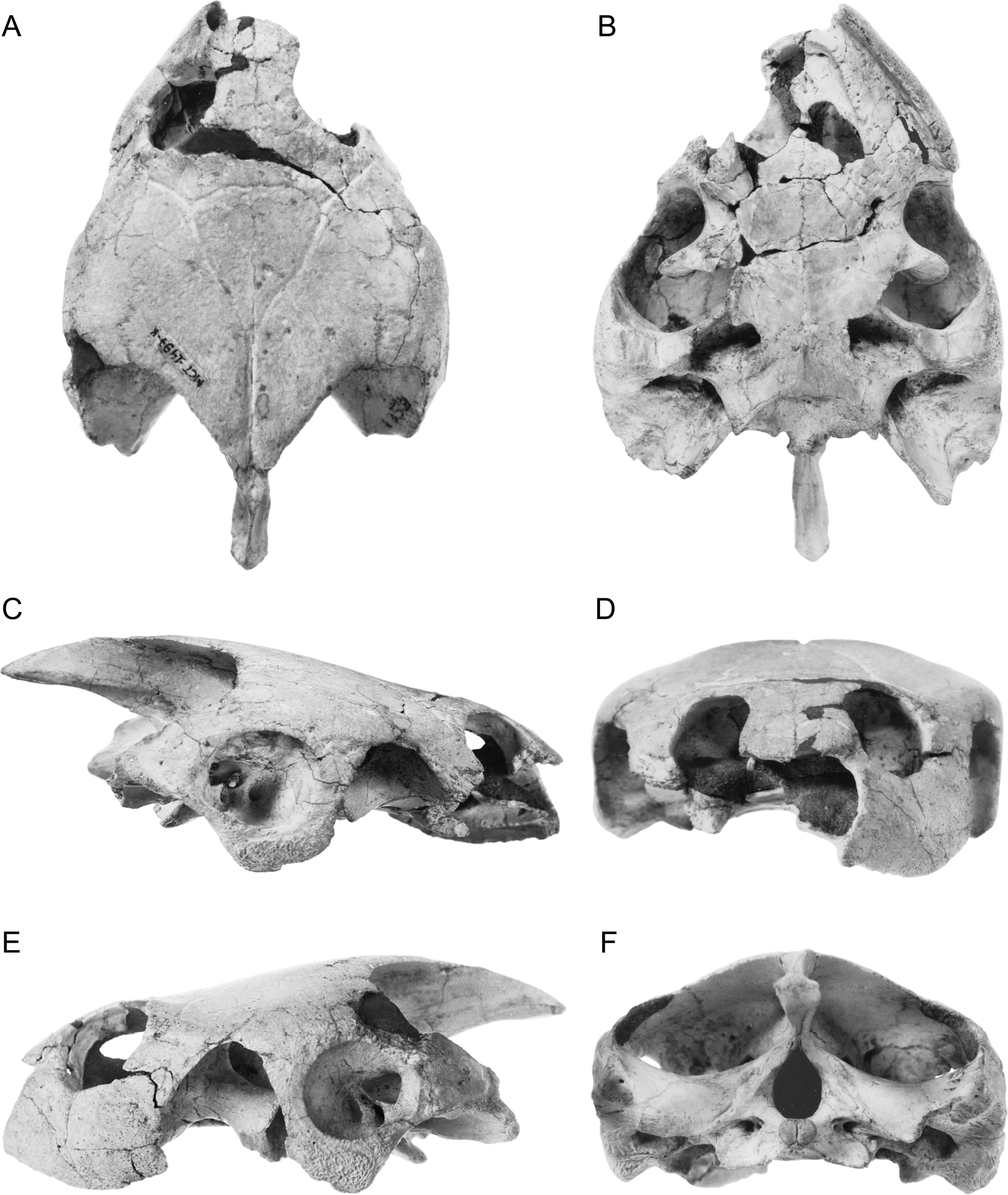
Fig. 7
Bauruemys elegans (Suárez,1969a). Partially restored skull based on DGM MCT 1492-R, DGM uncat A, MCZ 4123. A, dorsal; B, ventral; C, lateral. [A. Venjara, del.]
Fig. 8
Bauruemys elegans (Suárez, 1969a). Partially restored ventral view based on DGM MCT 1492-R and MCZ 4123. [A. Venjara, del.]
Fig. 9
Bauruemys elegans (Suárez,1969a). DGM MCT 1492-R, skull of holotyope. A, dorsal; B, ventral; C, right lateral; D, anterior; E, left lateral; F, posterior. [C. Facella del.]
Fig. 10
Bauruemys elegans (Suárez,1969a). DGM MCT 1492-R, skull of holotyope. A, dorsal; B, ventral; C, right lateral; D, anterior; E, left lateral; F, posterior. [C. Facella, del.]
Fig. 11
Bauruemys elegans (Suárez,1969a). DGM MCT 1753-R. A, dorsal; B, ventral; C, right lateral; D, anterior; E, left lateral; F, posterior. [M. Vabulas, del.]
Fig. 12
Bauruemys elegans (Suárez,1969a). DGM MCT 1753-R. A, dorsal; B, ventral; C, right lateral; D, anterior; E, left lateral; F, posterior. [M. Vabulas, del.]
Fig. 13
Bauruemys elegans (Suárez, 1969a). MCZ 4123. A, dorsal; B, ventral; C, right lateral; D, anterior; E, left lateral; F, posterior. [A. Venjara, del.]
Fig. 14
Bauruemys elegans (Suárez, 1969a). MCZ 4123. A, dorsal; B, ventral; C, right lateral; D, anterior; E, left lateral; F, posterior. [A. Venjara, del.]
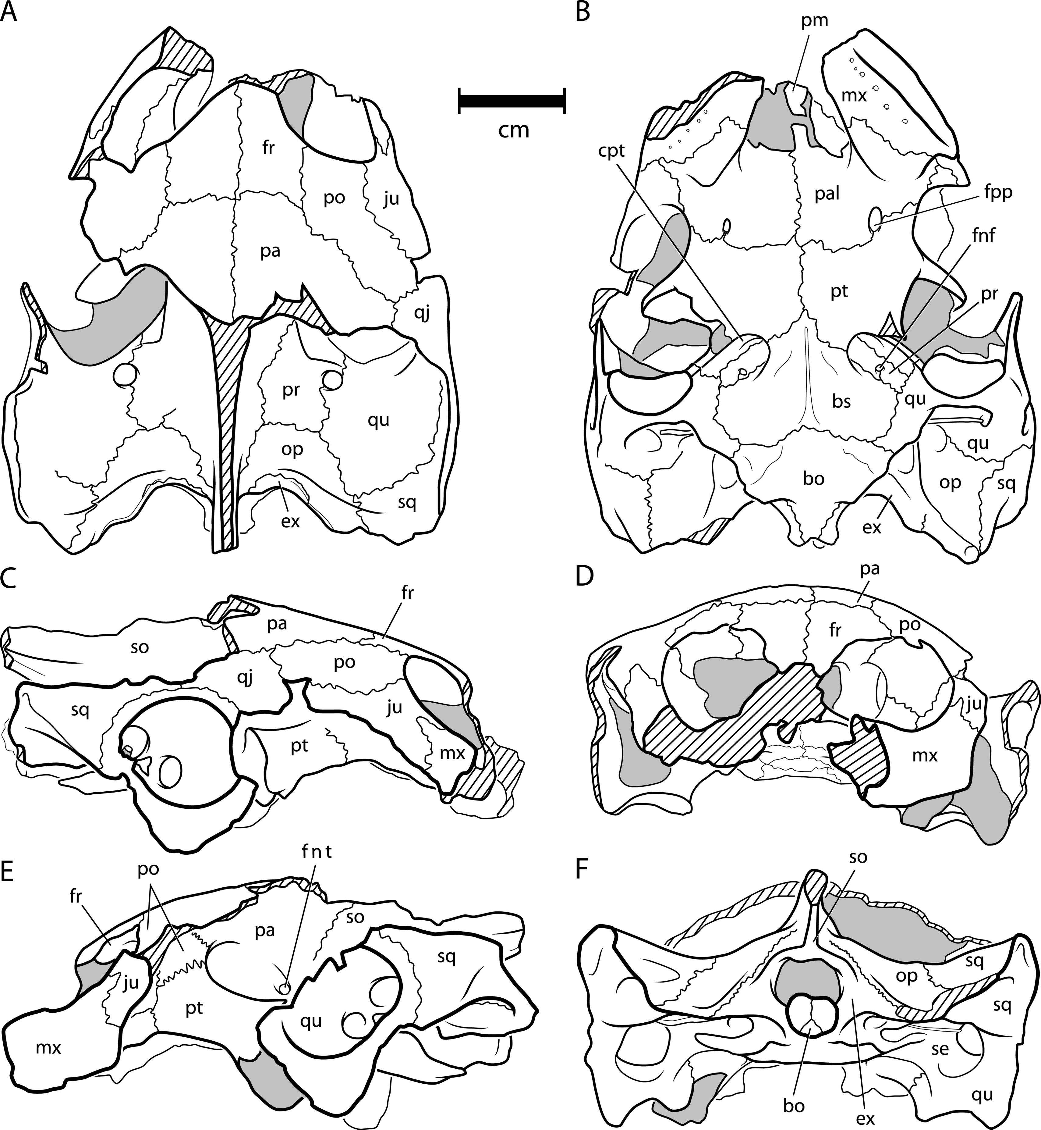
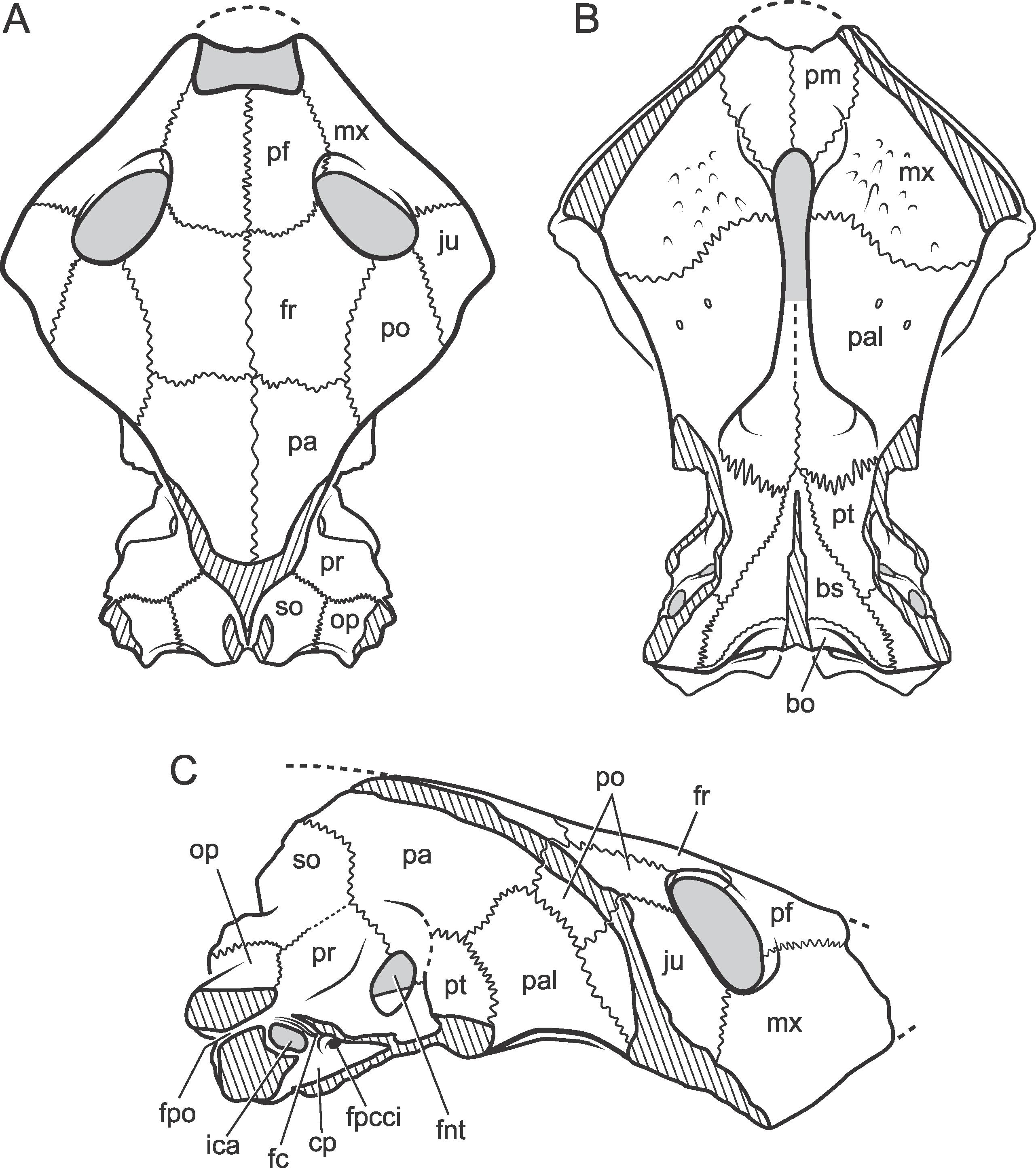
Fig. 15
Peiropemys mezzalirai, n. gen. et sp. Partially restored skull of holotype, DGM MCT 1497-R. A, dorsal; B, ventral; C, occipital; D, lateral. [P. Sloan, del.]
Fig. 16
Peiropemys mezzalirai, n. gen. et sp. Partially restored ventral view of skull DGM MCT 1497-R. [V. Storfer, del.]
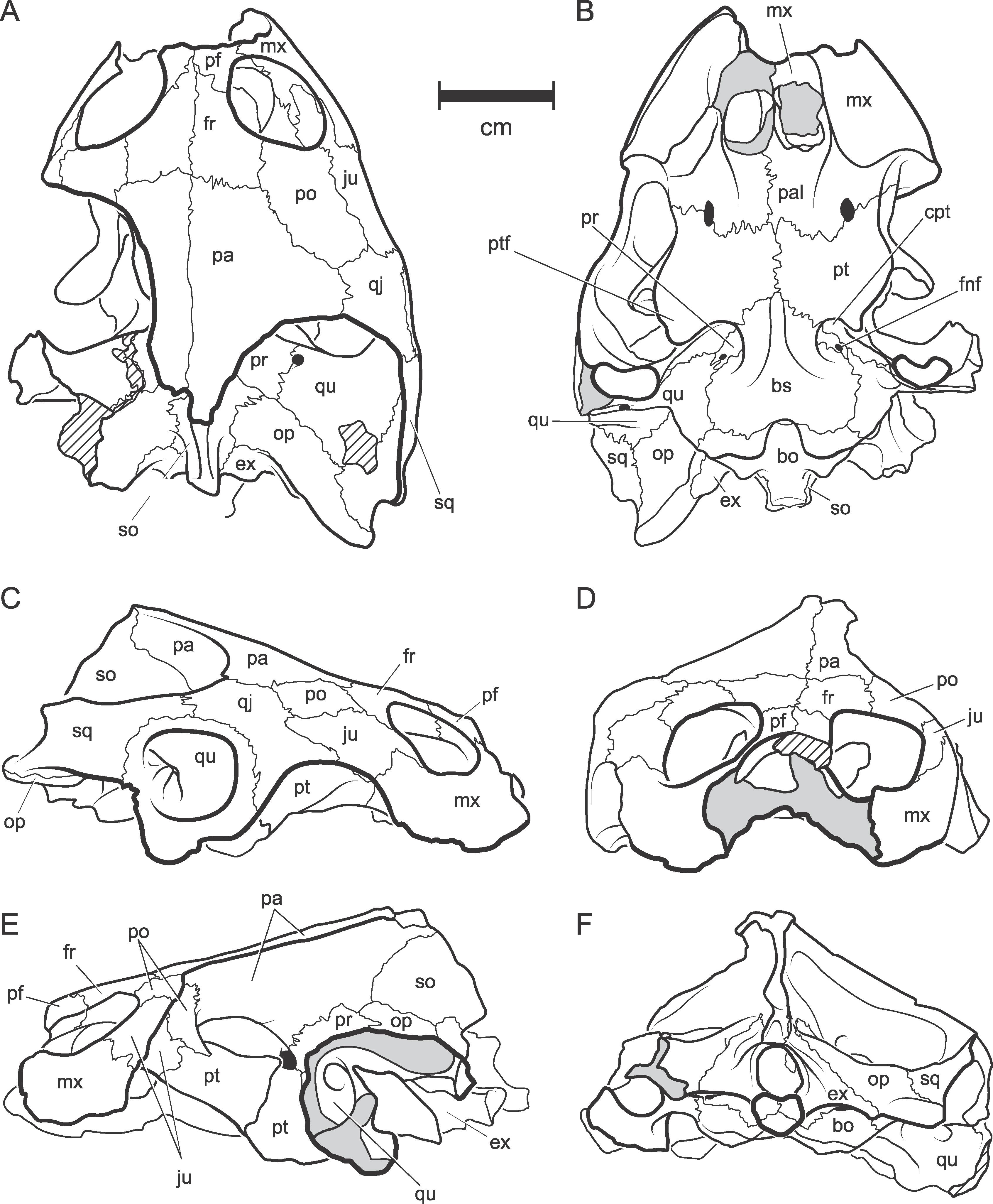
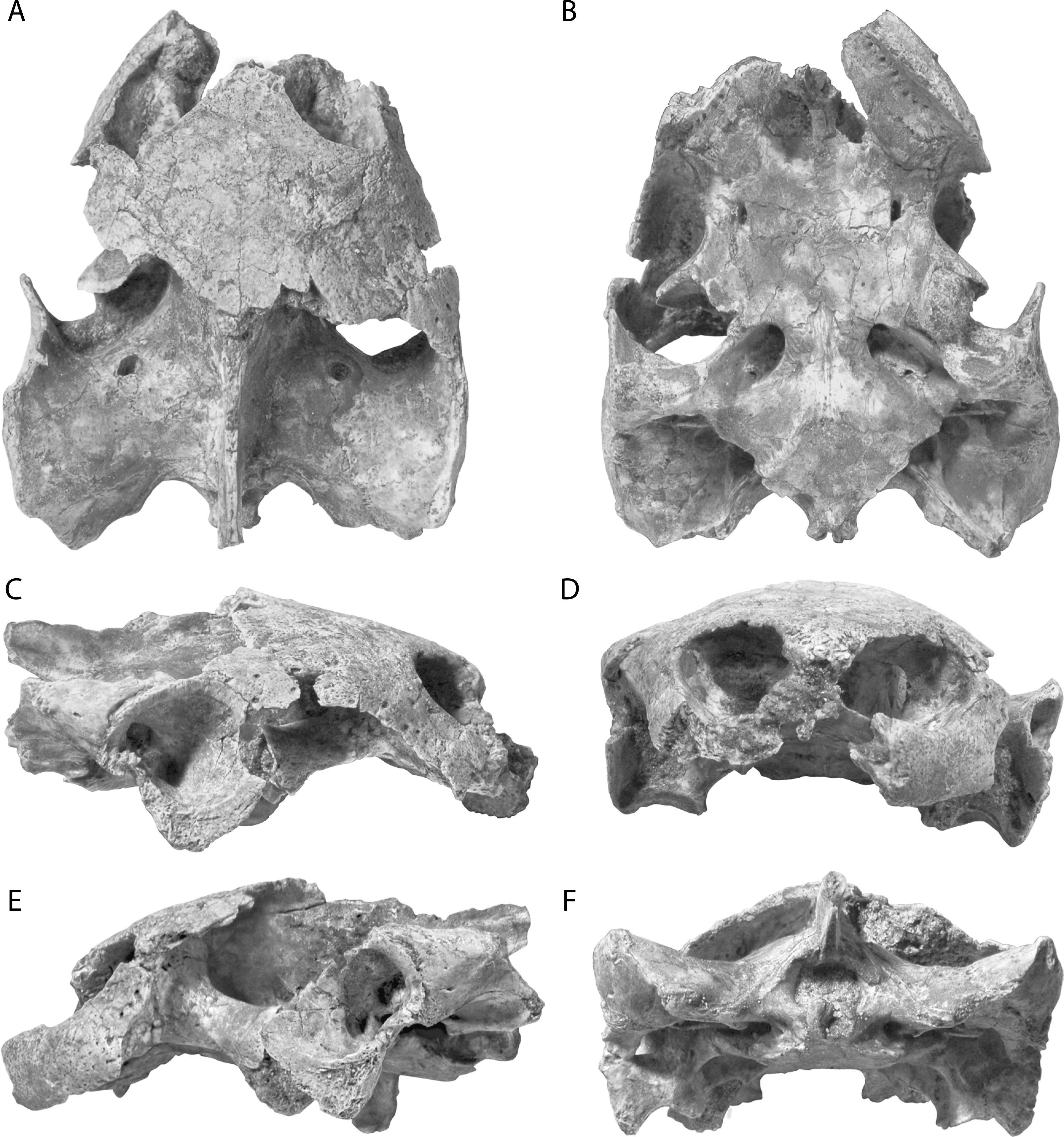
Fig. 17
Peiropemys mezzalirai, n. gen. et sp. DGM MCT 1497-R. Skull. A, dorsal; B, ventral; C, right lateral; D, anterior; E, left lateral; F, posterior. [P. Sloan, G. Giardina, del.]
Fig. 18
Peiropemys mezzalirai, n. gen. et sp. DGM MCT 1497-R. Skull. A, dorsal; B, ventral; C, right lateral; D, anterior; E, left lateral; F, posterior. [C. Facella, del.]
Fig. 19
Lapparentemys vilavilensis (Broin, 1971), n. gen. Partially restored skull based on AMNH 14444 and WUS 2160. A, dorsal; B, ventral; C, lateral. [F. Ippolito, del.]
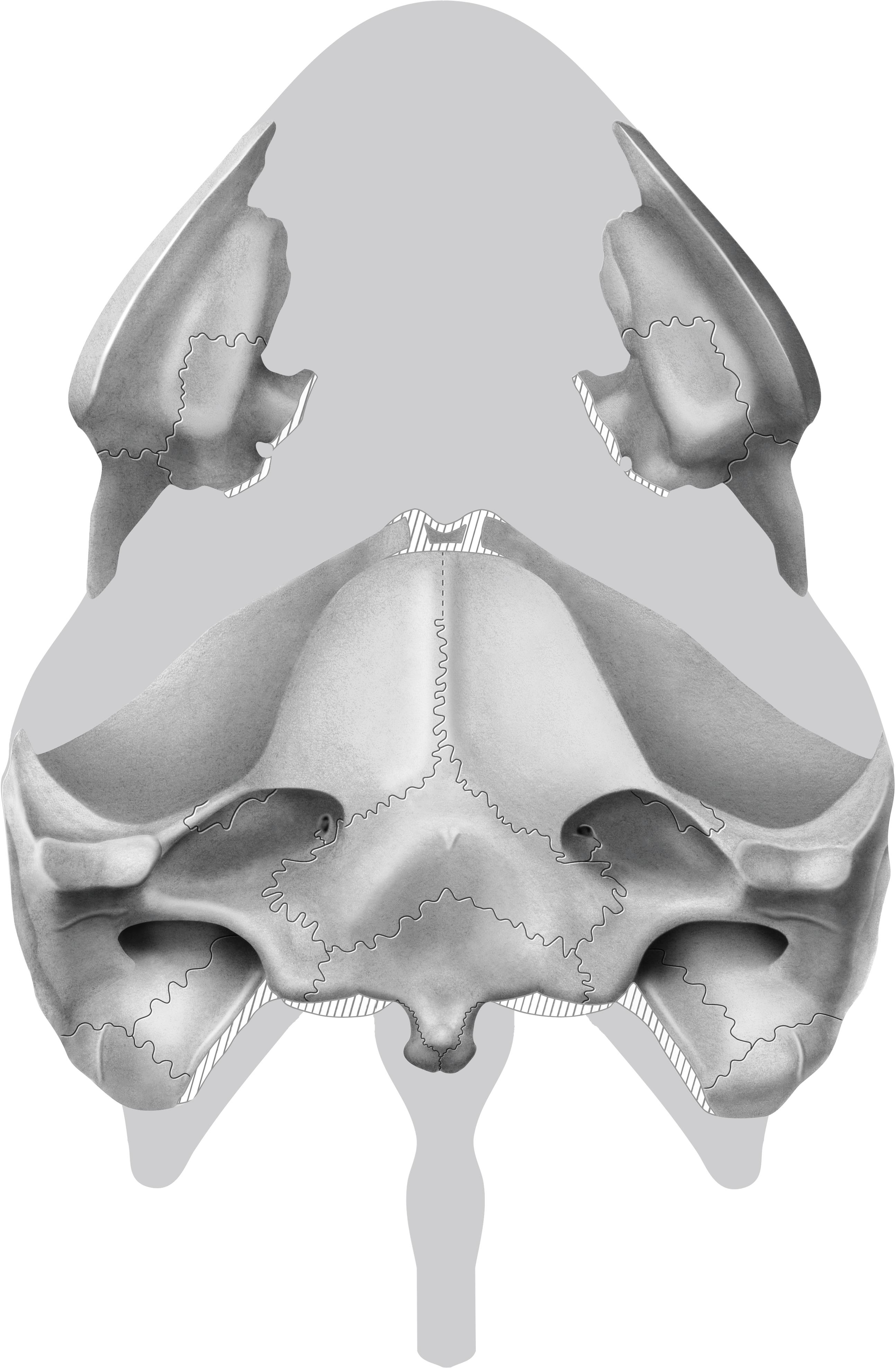
Fig. 20
Lapparentemys vilavilensis (Broin, 1971), n. gen. Partially restored ventral view of skull based on AMNH 14444. [T. Tarpley, del.]
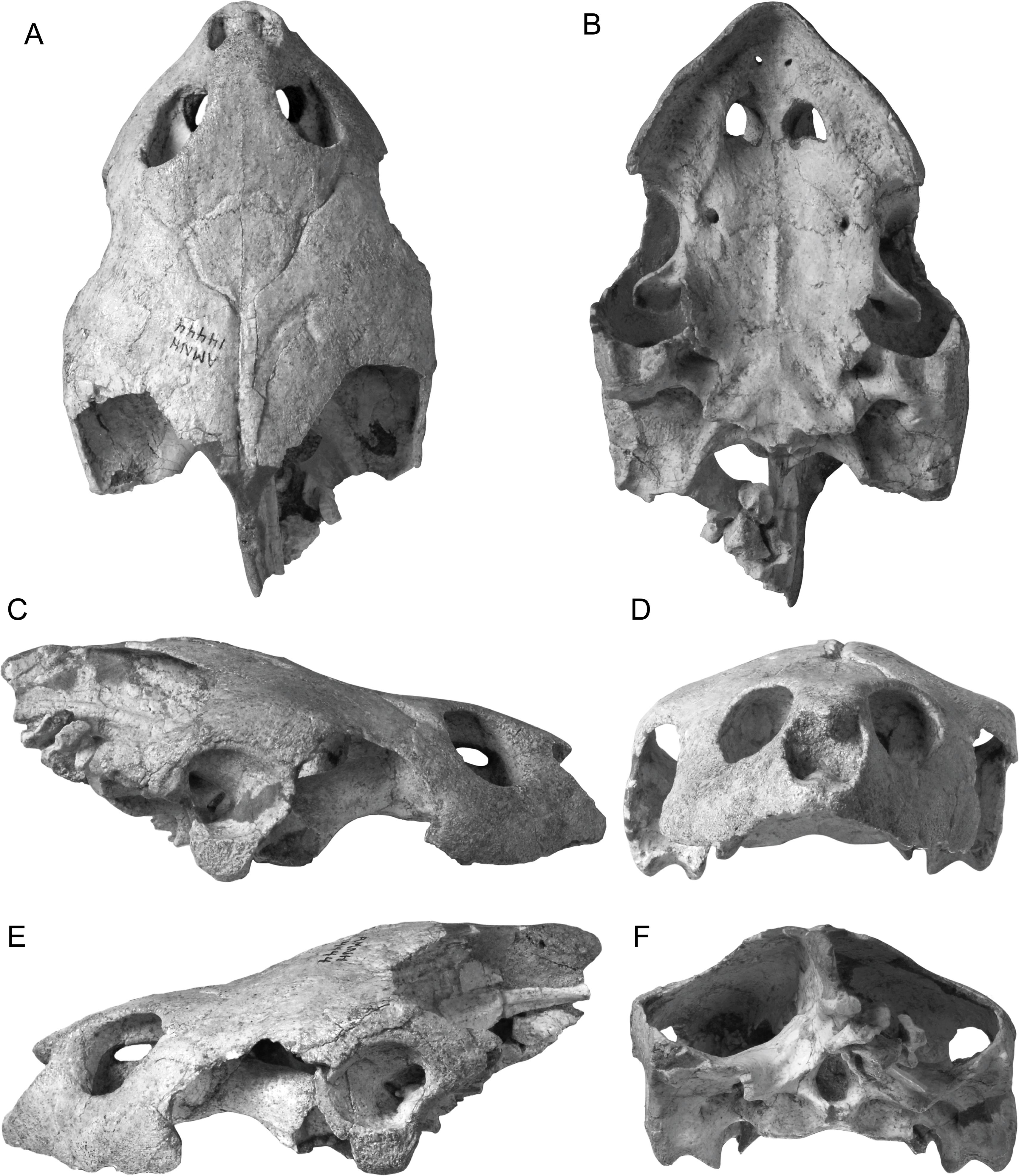
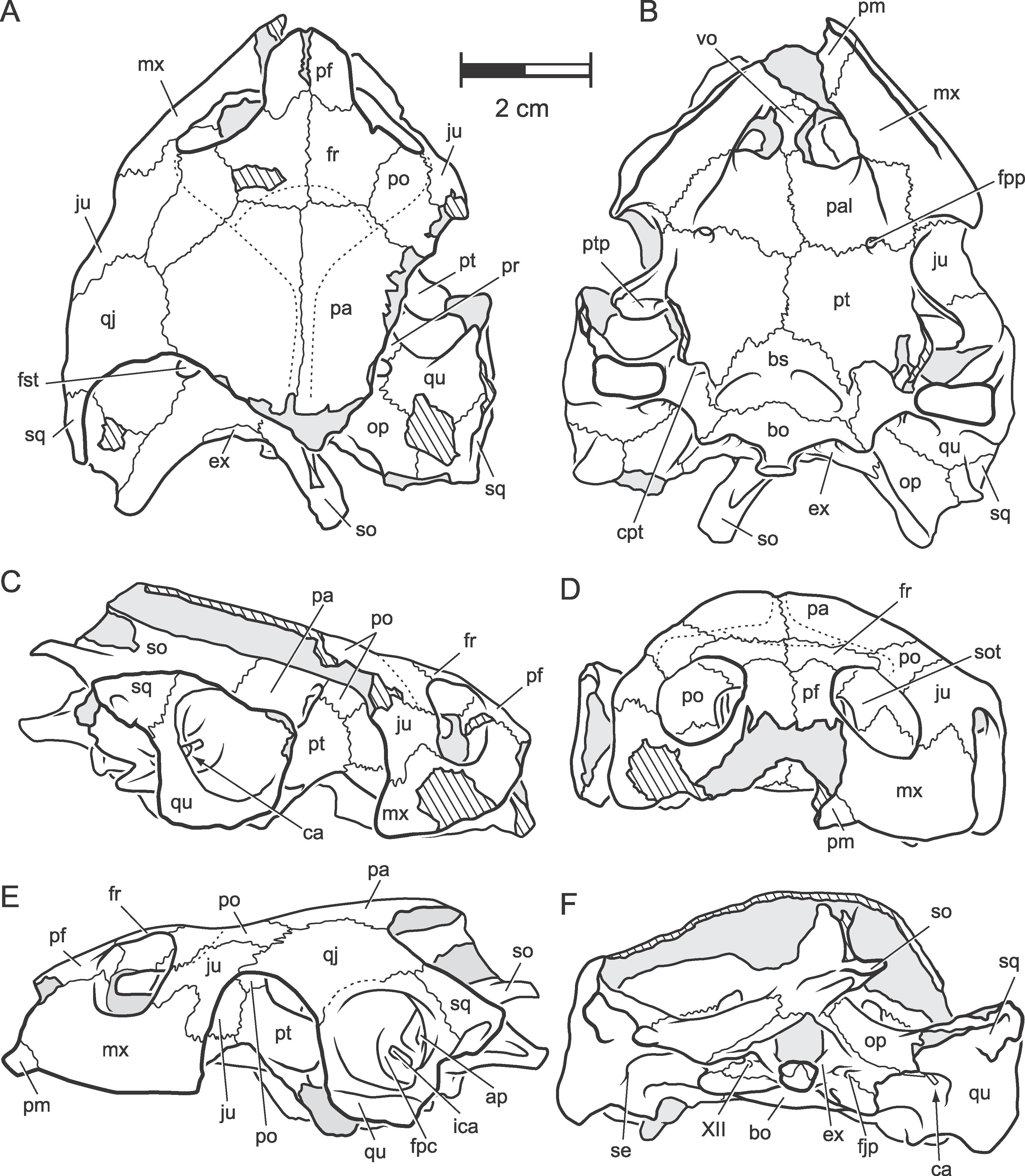
Fig. 21
Lapparentemys vilavilensis (Broin, 1971), n. gen. Skull. AMNH 14444 . A, dorsal; B, ventral; C, right lateral; D, anterior; E, left lateral; F, posterior. [F. Ippolito, C. Facella, del.]
Fig. 22
Lapparentemys vilavilensis (Broin, 1971), n. gen. Skull. AMNH 14444. A, dorsal; B, ventral; C, right lateral; D, anterior; E, left lateral; F, posterior. [F. Ippolito, del.]
Fig. 23
Lapparentemys vilavilensis (Broin, 1971), n. gen. Partially restored skull based on WUS 2160. A, dorsal; B, ventral; C, lateral. [C. Wilson, del.]
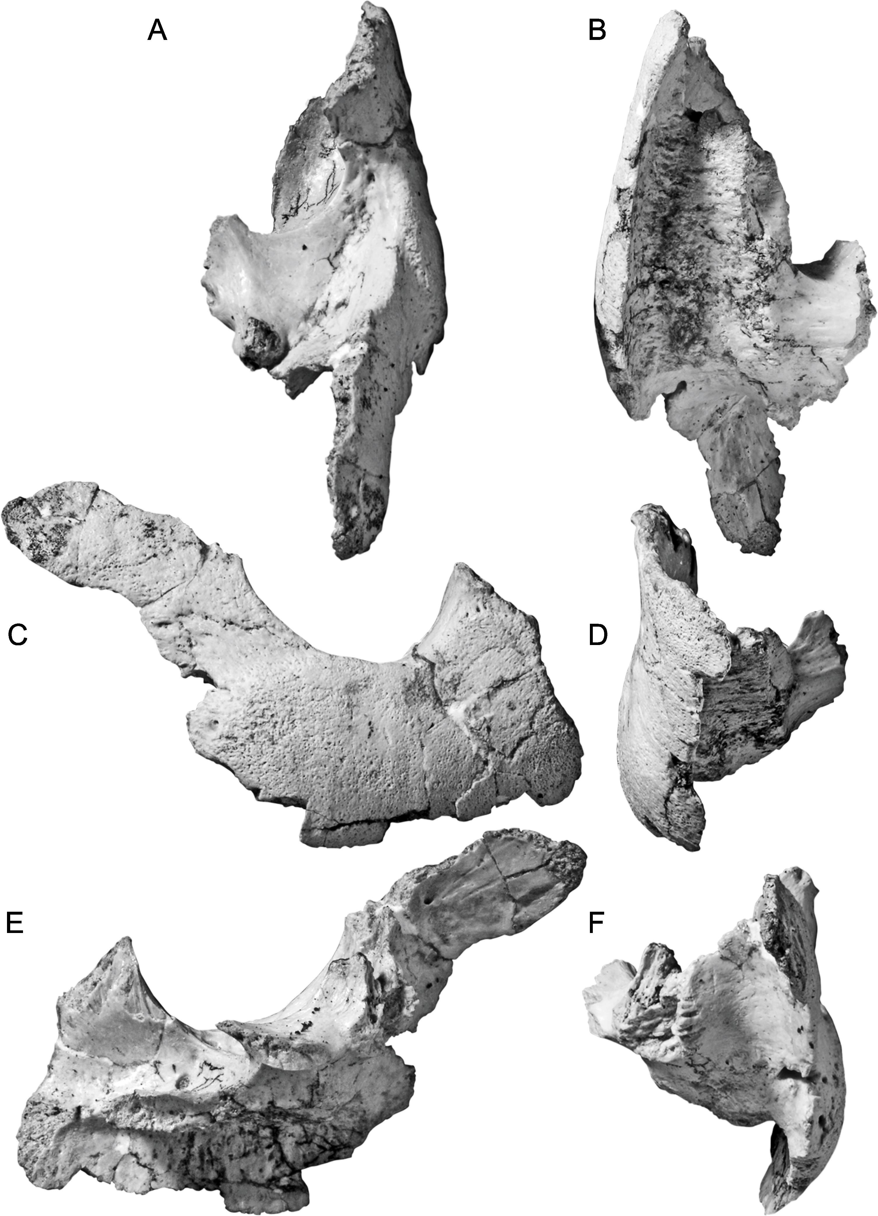
Fig. 24
Lapparentemys vilavilensis (Broin, 1971), n. gen. Skull WUS 2160. A, dorsal; B, ventral; C, right lateral; D, anterior; E, left lateral; F, posterior. [F. Ippolito, C. Facella, del.]
Fig. 25
Lapparentemys vilavilensis (Broin, 1971), n. gen. Skull WUS 2160. A, dorsal; B, ventral; C, right lateral; D, anterior; E, left lateral; F, posterior. [C. Wilson, del.]
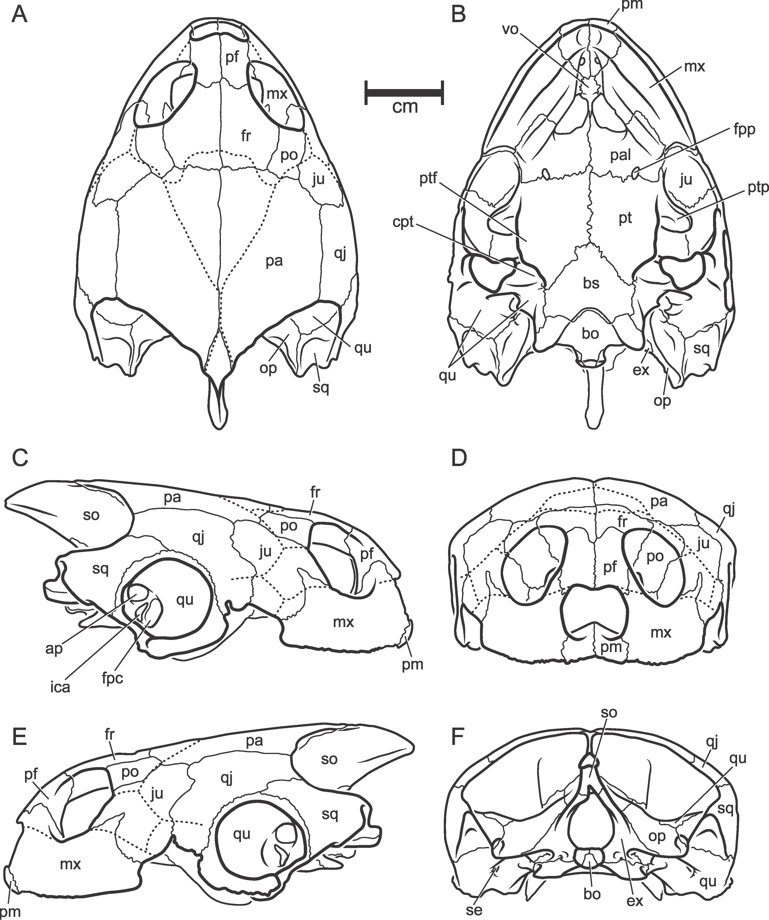
Fig. 26
Pricemys caiera, n. gen. et sp. Partially restored skull based on DGM MCT 1498-R. A, dorsal; B, ventral; C, lateral. [M. Beveridge, del.]
Fig. 27
Pricemys caiera, n. gen. et sp. Partially restored ventral view of skull based on DGM MCT 1498-R. [M. Beveridge, del.]
Fig. 28
Pricemys caiera, n. gen. et sp. DGM MCT 1498-R. Braincase moiety. A, dorsal; B, ventral; C, right lateral; D, anterior; E, left lateral; F, posterior. [C. Facella, del.]
Fig. 29
Pricemys caiera, n. gen. et sp. DGM MCT 1498-R. Braincase moiety. A, dorsal; B, ventral; C, right lateral; D, anterior; E, left lateral; F, posterior. [C. Facella, del.]
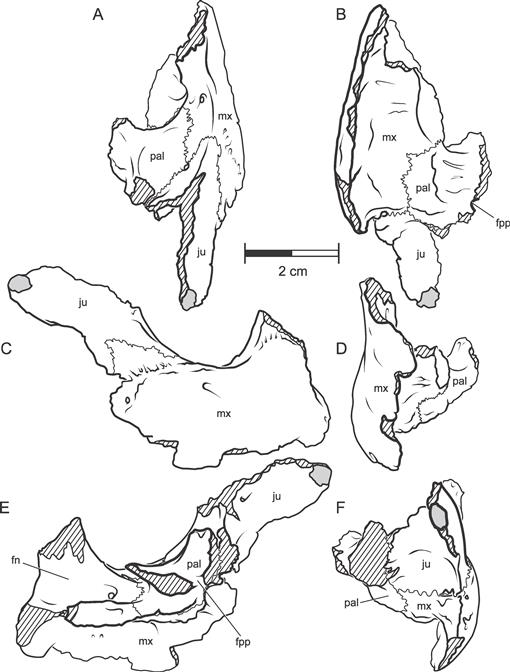
Fig. 30
Pricemys caiera, n. gen. et sp. DGM MCT 1498-R. Jugal-maxilla-palatine moiety. A, dorsal; B, ventral; C, right lateral; D, anterior; E, left lateral; F, posterior. [C. Facella, del.]
Fig. 31
Pricemys caiera, n. gen. et sp. DGM MCT 1498-R. Jugal-maxilla-palatine moiety. A, dorsal; B, ventral; C, right lateral; D, anterior; E, left lateral; F, posterior. [J. Sharkey, del.]
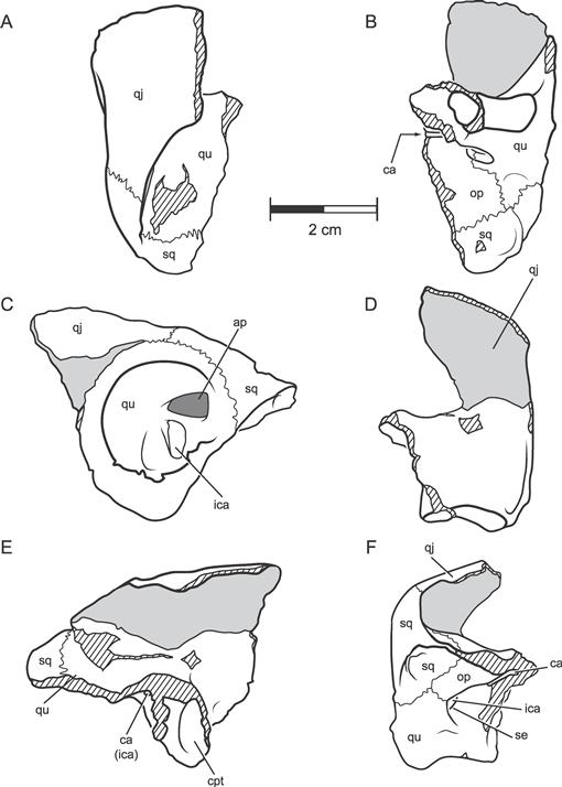
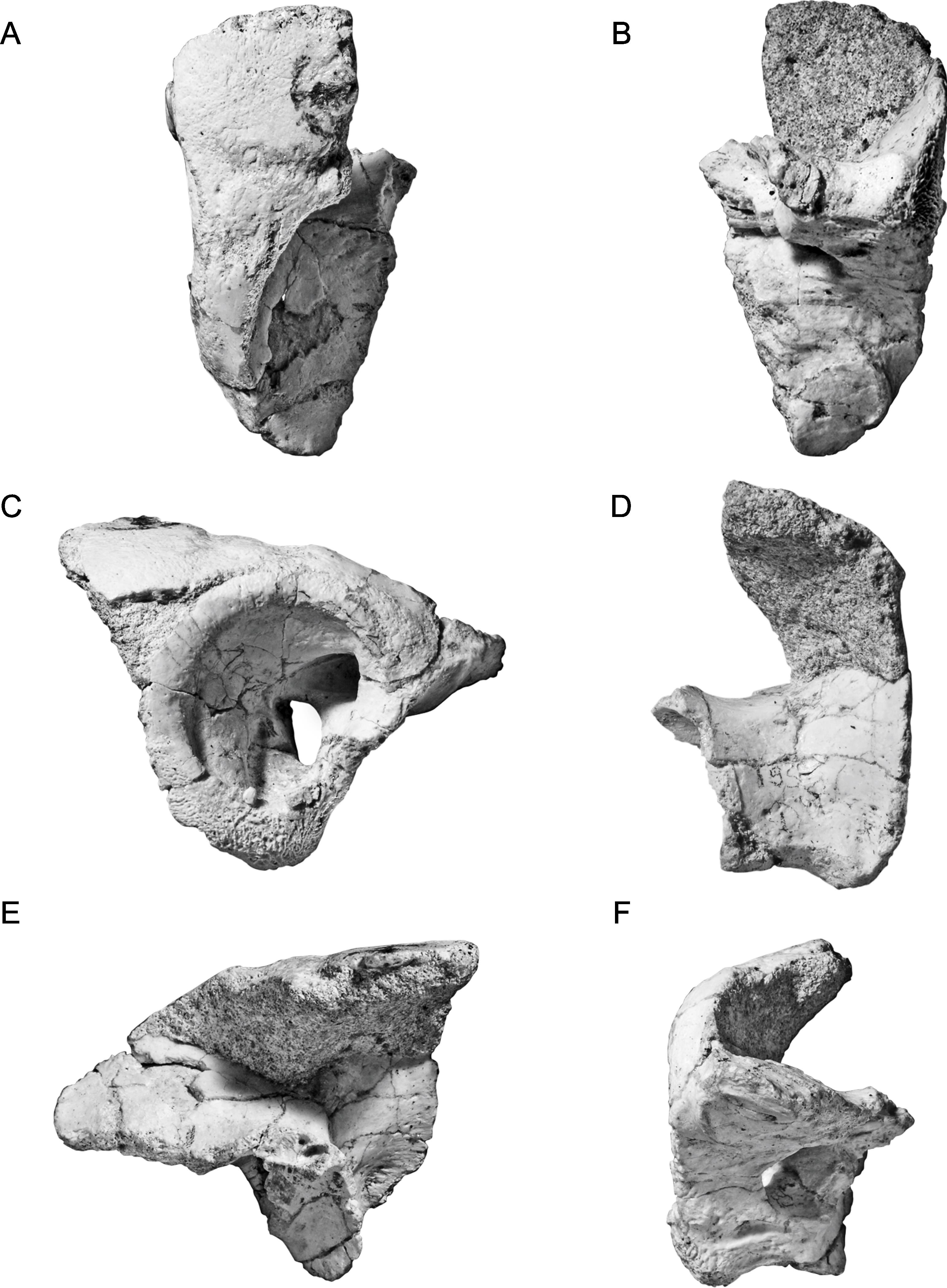
Fig. 32
Pricemys caiera, n. gen. et sp. DGM MCT 1498-R. Left quadrate moiety. A, dorsal; B, ventral; C, left lateral; D, anterior; E, medial; F, posterior. [C. Facella, del.]
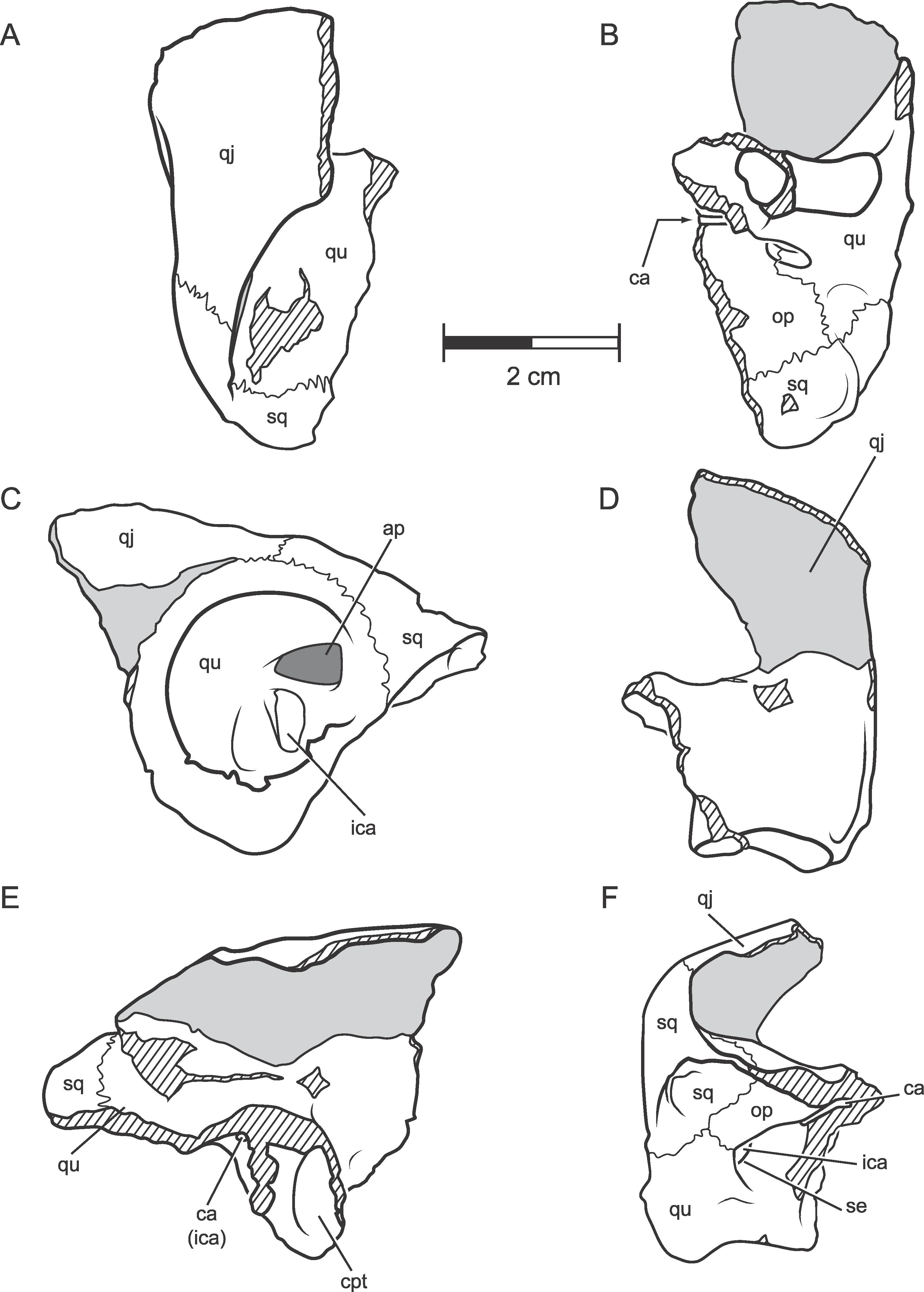
Fig. 33
Pricemys caiera, n. gen. et sp. DGM MCT 1498-R. Left quadrate moiety. A, dorsal; B, ventral; C, left lateral; D, anterior; E, medial; F, posterior. [C. Facella, del.]
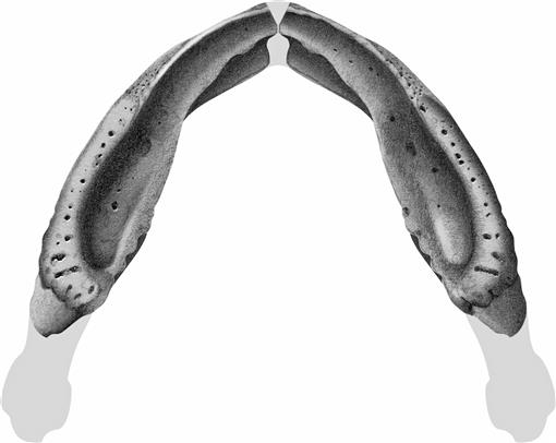
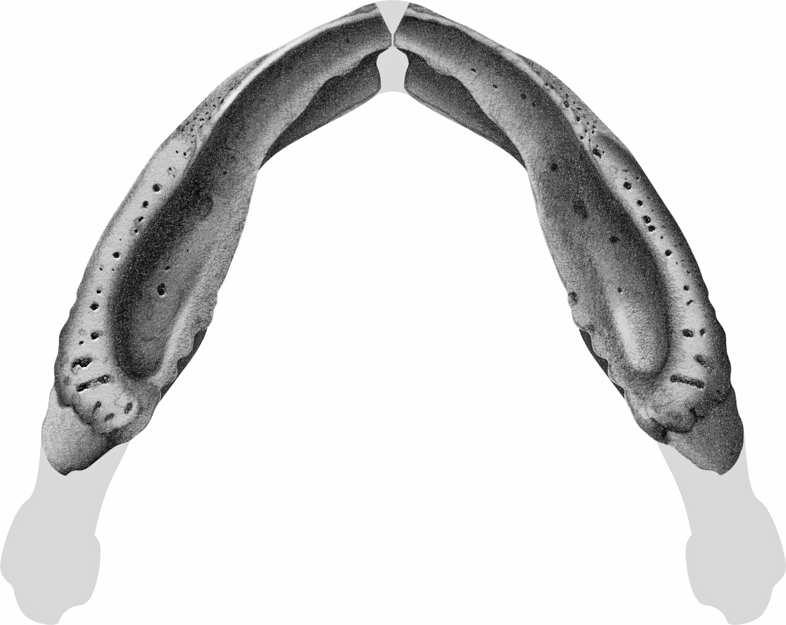
Fig. 34
?Pricemys caiera, n. gen. et sp. DGM MCT ? R. Dorsal view of lower jaw, right ramus reversed. [F. Ippolito, del.]
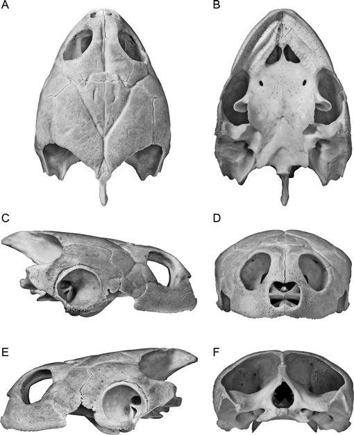
Fig. 35
Podocnemis vogli Müller, 1935. Skull, UF 39100. A, dorsal; B, ventral; C, right lateral; D, anterior; E, left lateral; F, posterior. [M. Vabulas, del.]
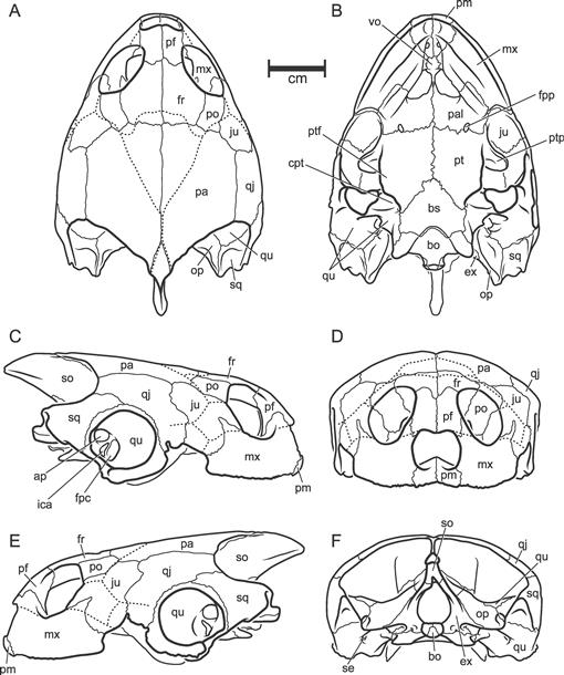
Fig. 36
Podocnemis vogli Müller, 1935. Skull, UF 39100. A, dorsal; B, ventral; C, right lateral; D, anterior; E, left lateral; F, posterior. [M. Vabulas, del.]
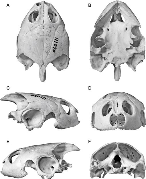
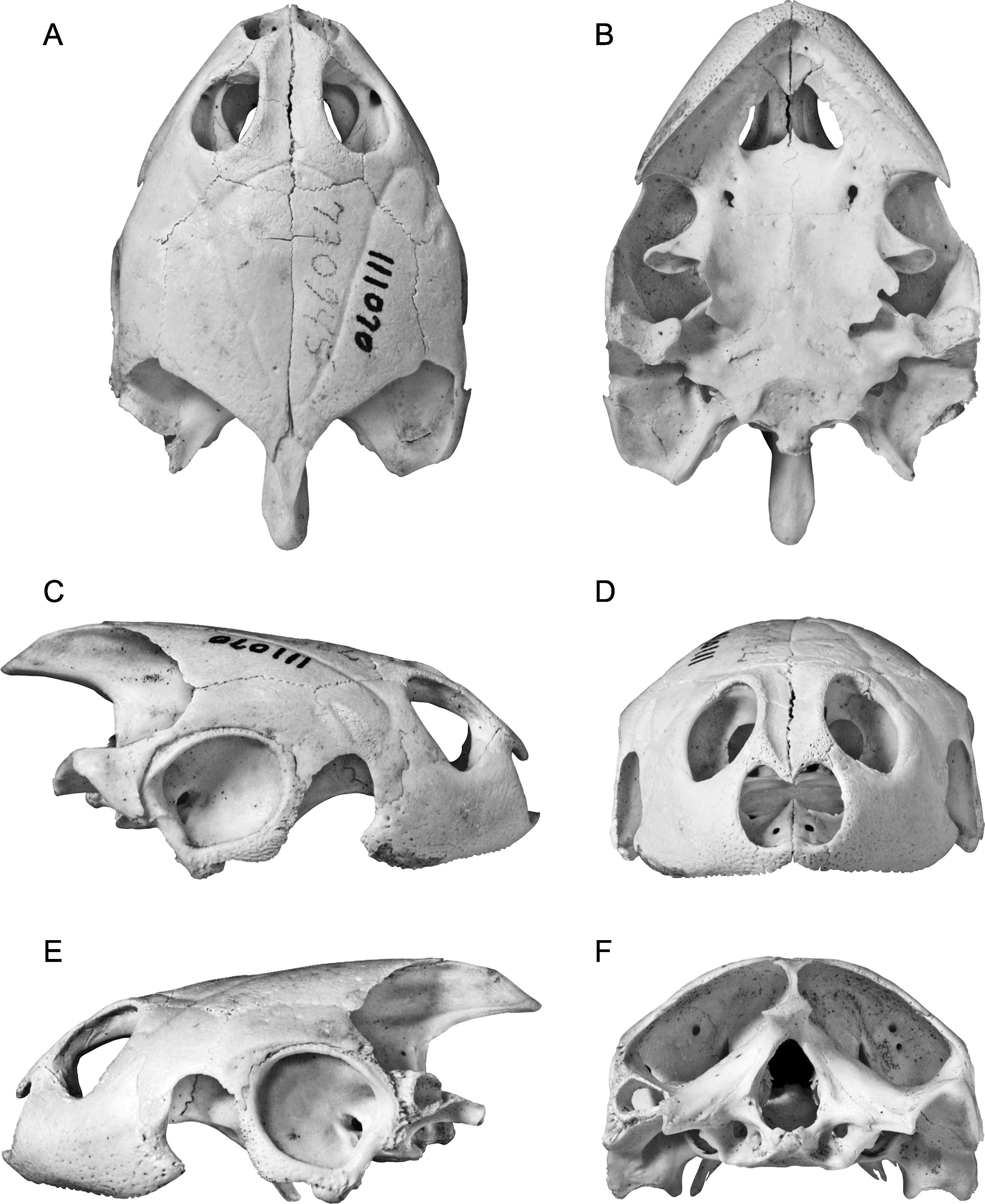
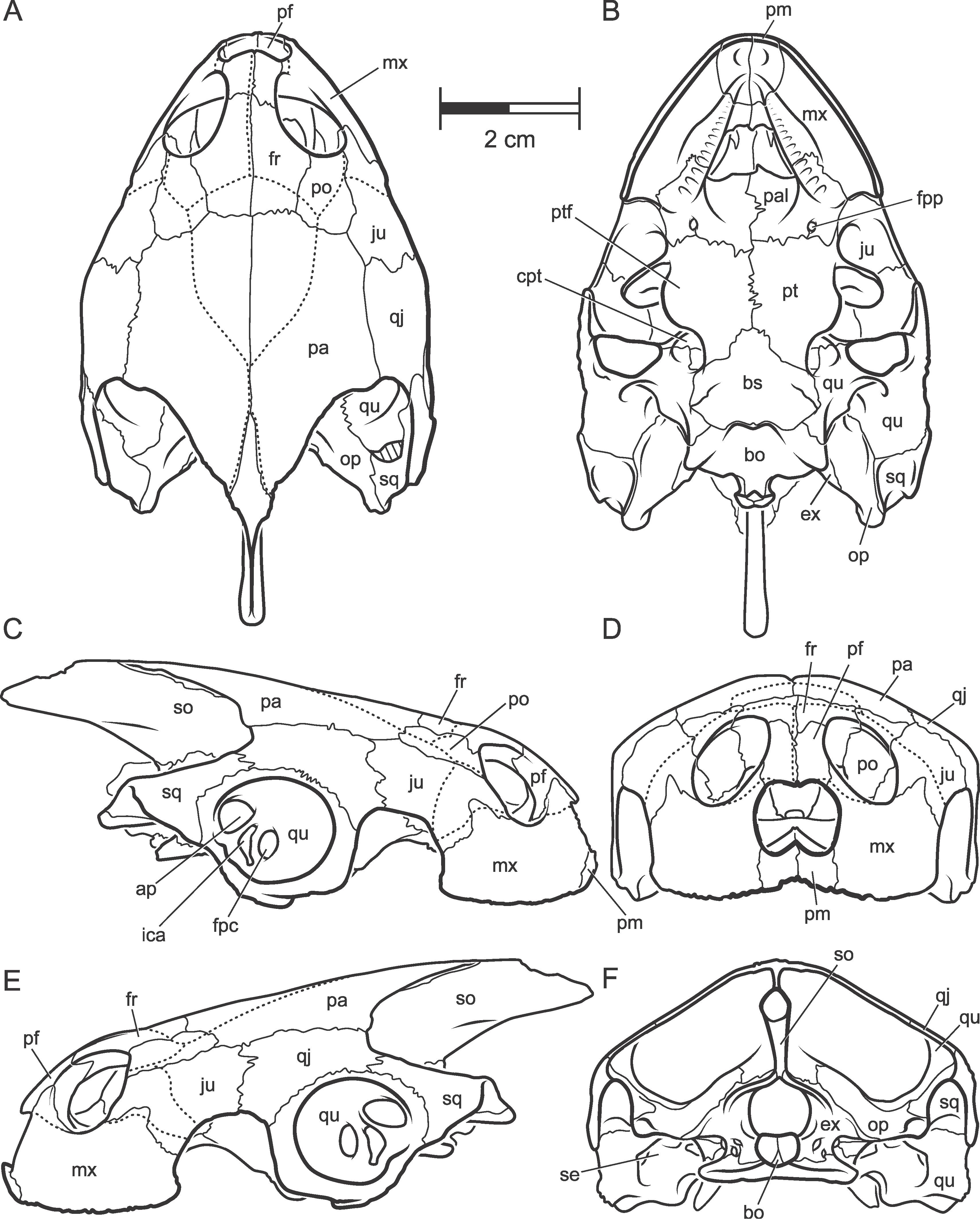
Fig. 37
Podocnemis sextuberculata Cornalia, 1849. Skull, AMNH 111070 A, dorsal; B, ventral; C, right lateral; D, anterior; E, left lateral; F, posterior. [M. Vabulas, del.]
Fig. 38
Podocnemis sextuberculata Cornalia, 1849. Skull, AMNH 111070 A, dorsal; B, ventral; C, right lateral; D, anterior; E, left lateral; F, posterior. [M. Vabulas, del.]
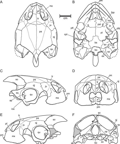
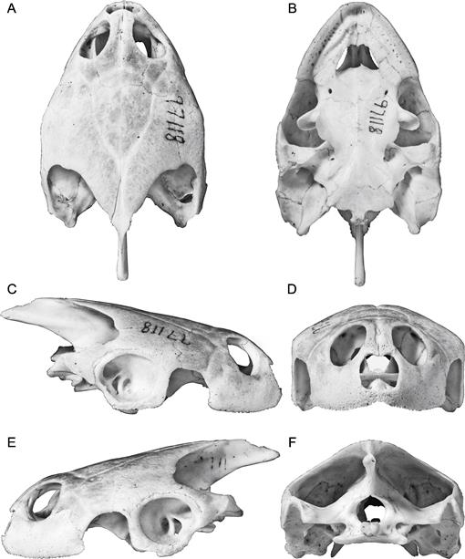
Fig. 39
Podocnemis unifilis Troschel, 1848. Skull, AMNH 97118. A, dorsal; B, ventral; C, right lateral; D, anterior; E, left lateral; F, posterior. [C. Facella, del.]
Fig. 40
Podocnemis unifilis Troschel, 1848. Skull, AMNH 97118. A, dorsal; B, ventral; C, right lateral; D, anterior; E, left lateral; F, posterior. [C. Facella, del.]
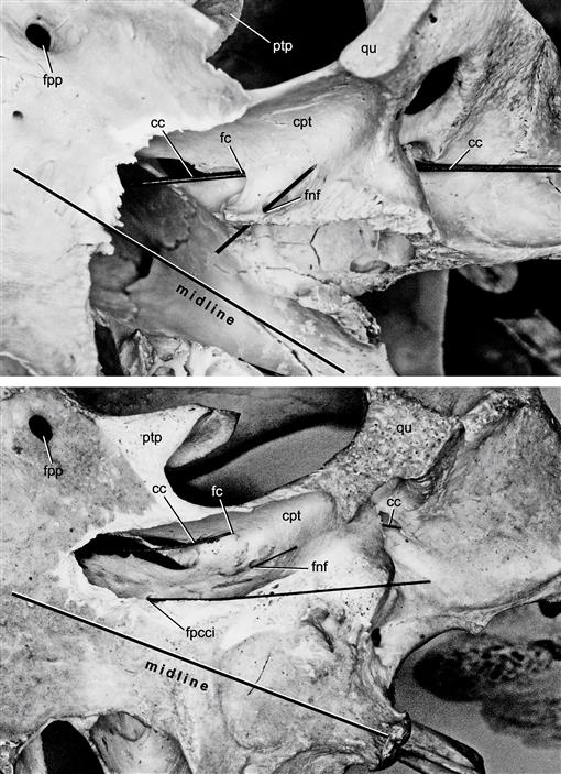
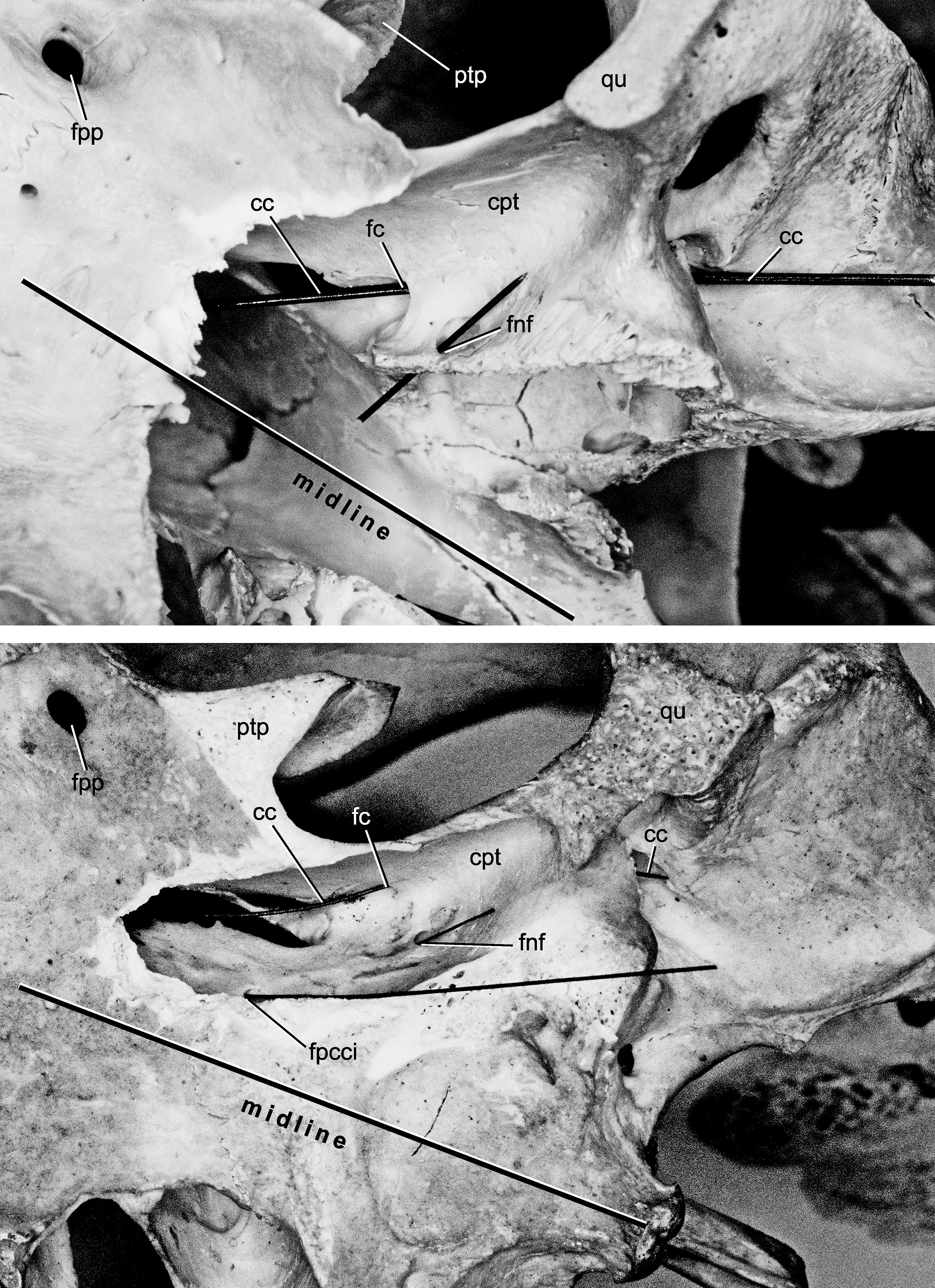
Fig. 41
Peltocephalus dumerilianus Schweigger, 1812. Ventral views of left cavum pterygoidi prepared out to show foramina. Upper, AMNH Herp R163020; lower, NFWFL 338. In both views a probe lies in the canalis cavernosus and the foramen nervi facialis. [E. Gaffney, del.]
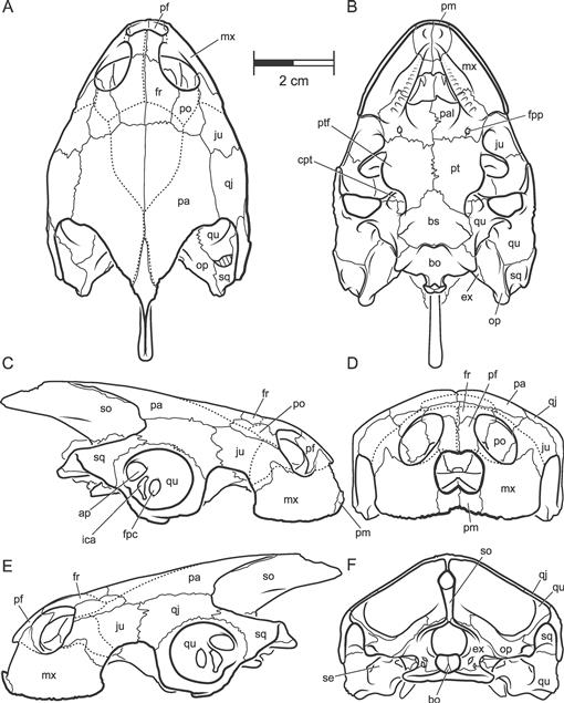
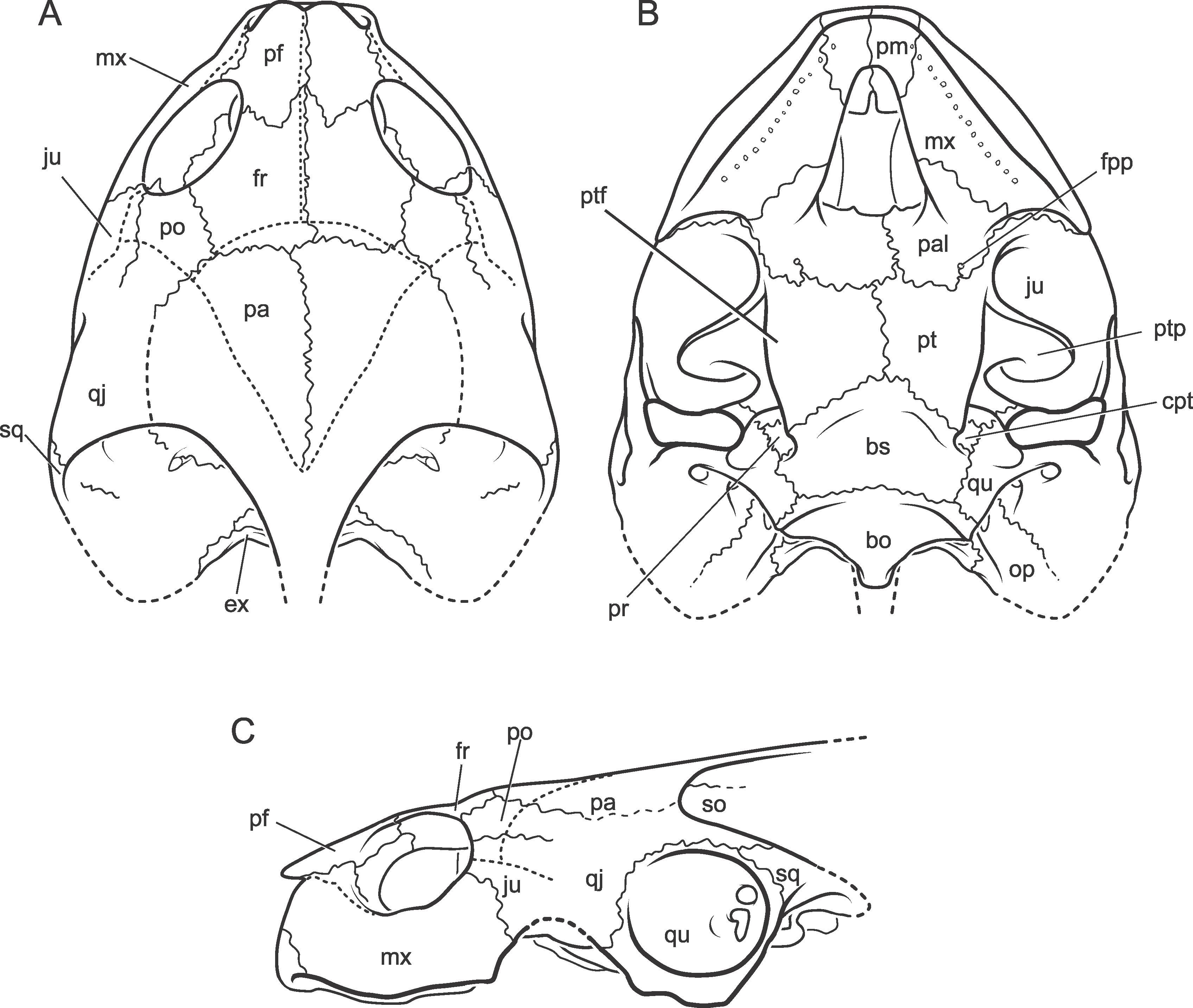
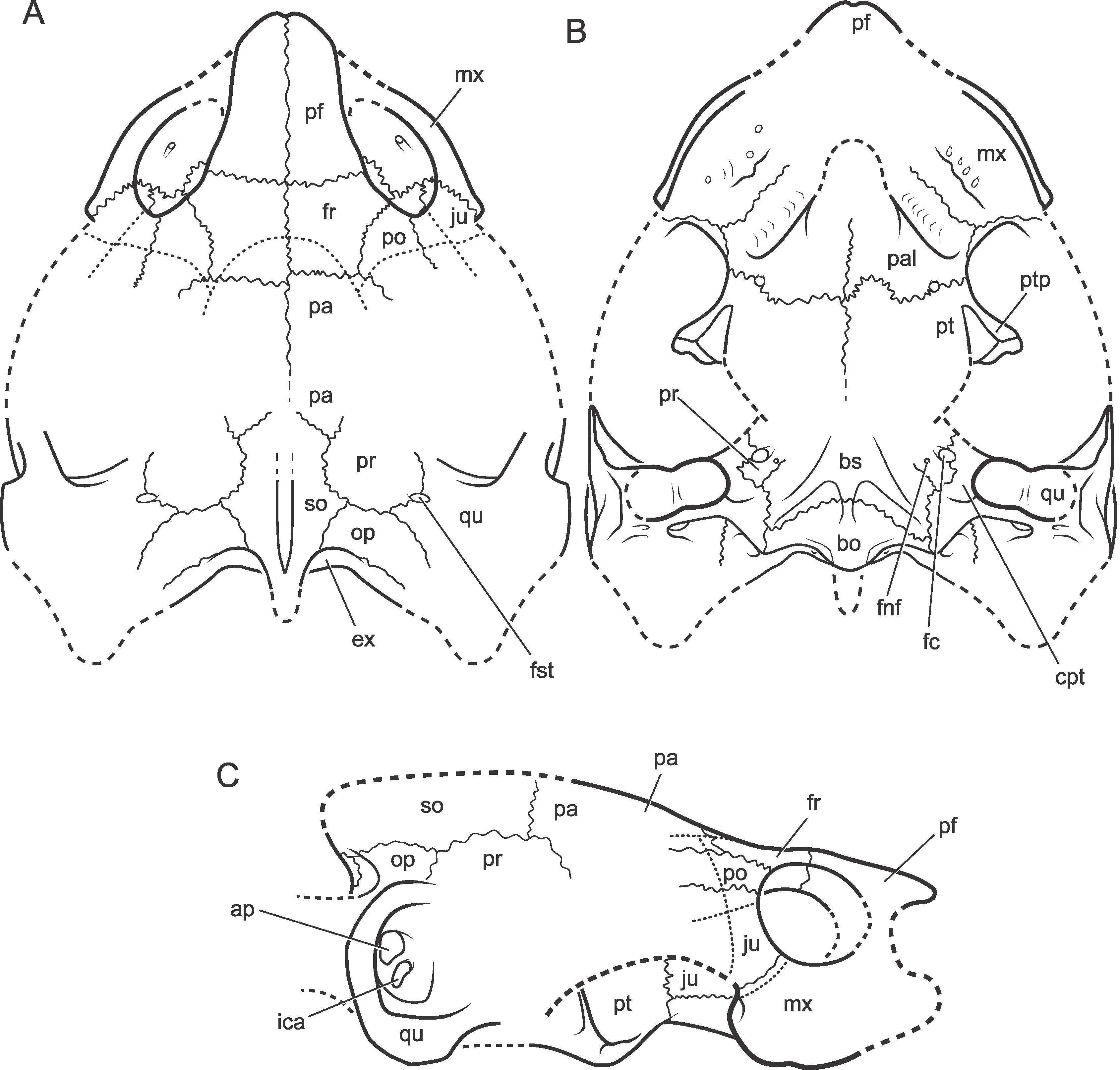
Fig. 42
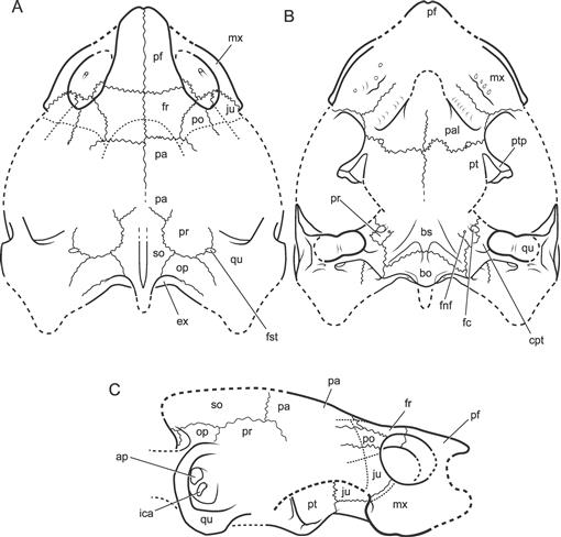
Neochelys fajumensis (Andrews, 1903). Partially restored skull based on DPC 3146. A, dorsal; B, ventral; C, lateral. [C. Facella, del.]
Fig. 43
Neochelys fajumensis (Andrews, 1903). Partially restored ventral view of skull of DPC 3146. [J. Lovell, del.]
Fig. 44
Neochelys fajumensis (Andrews, 1903). DPC 3146. Skull. A, dorsal; B, ventral; C, right lateral; D, anterior; E, left lateral; F, posterior. [C. Facella, del.]
Fig. 45
Neochelys fajumensis (Andrews, 1903). DPC 3146. A, dorsal; B, ventral; C, right lateral; D, anterior; E, left lateral; F, posterior. [C. Facella, del.]
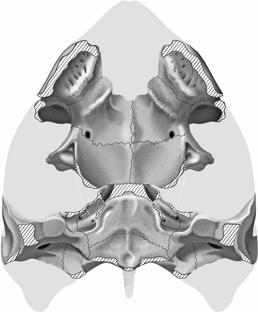
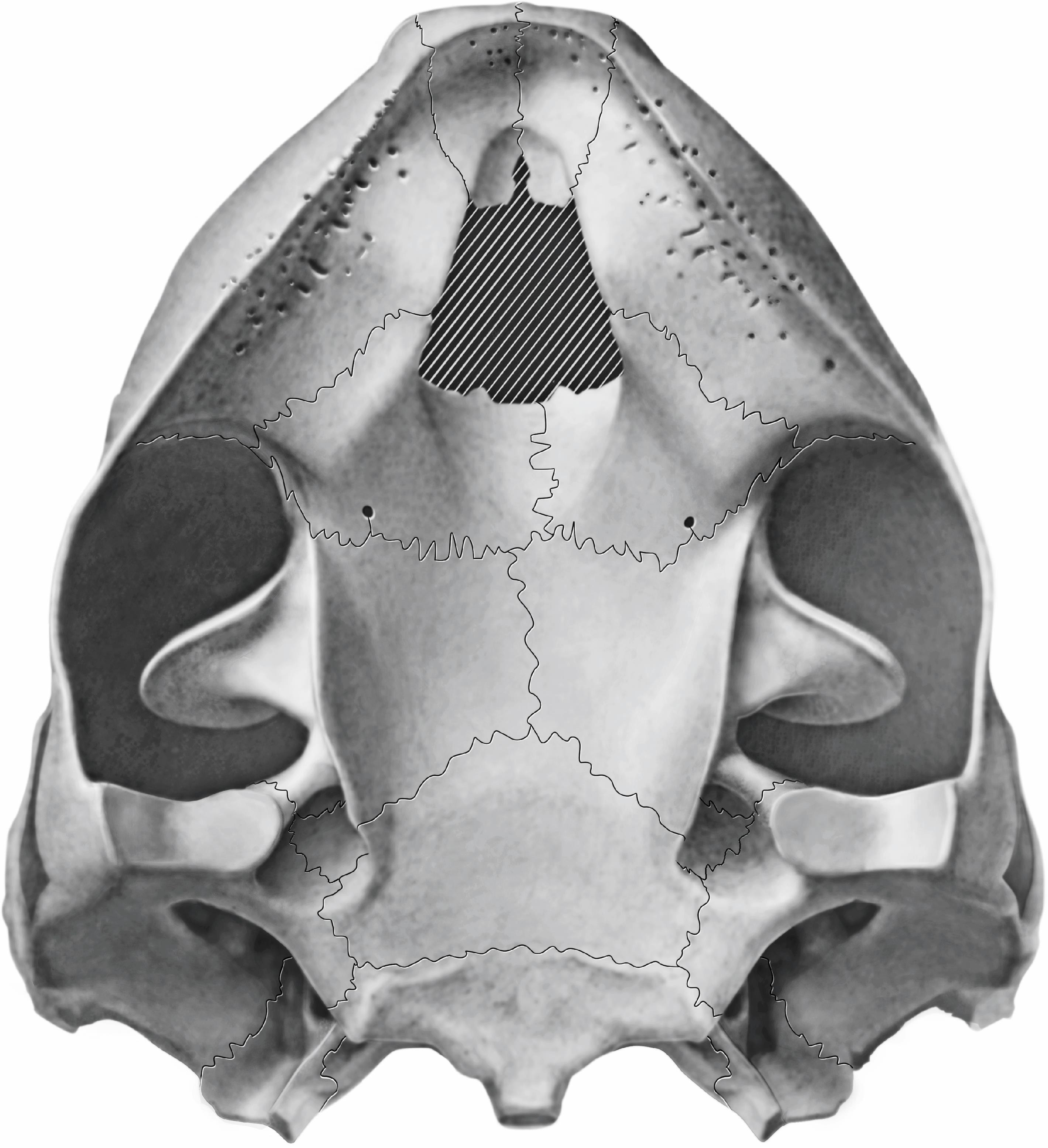
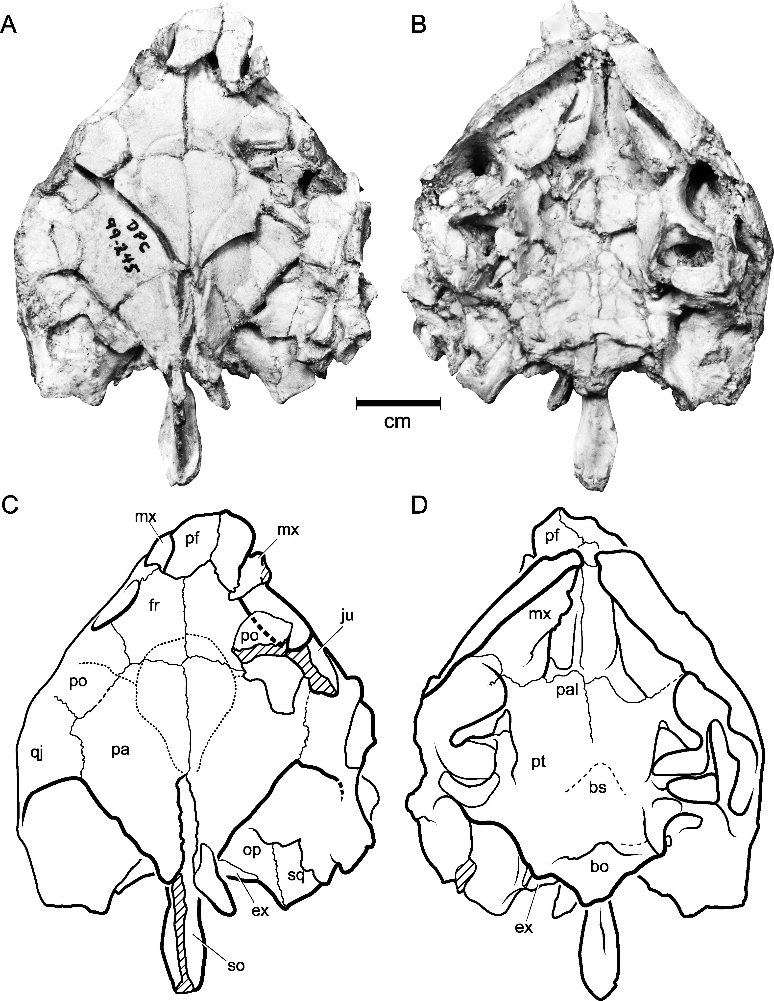
Fig. 46
Neochelys fajumensis (Andrews, 1903). DPC 99–245. Skull. A, C, dorsal views; B, D, ventral views.
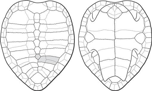
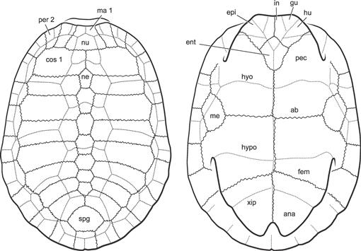
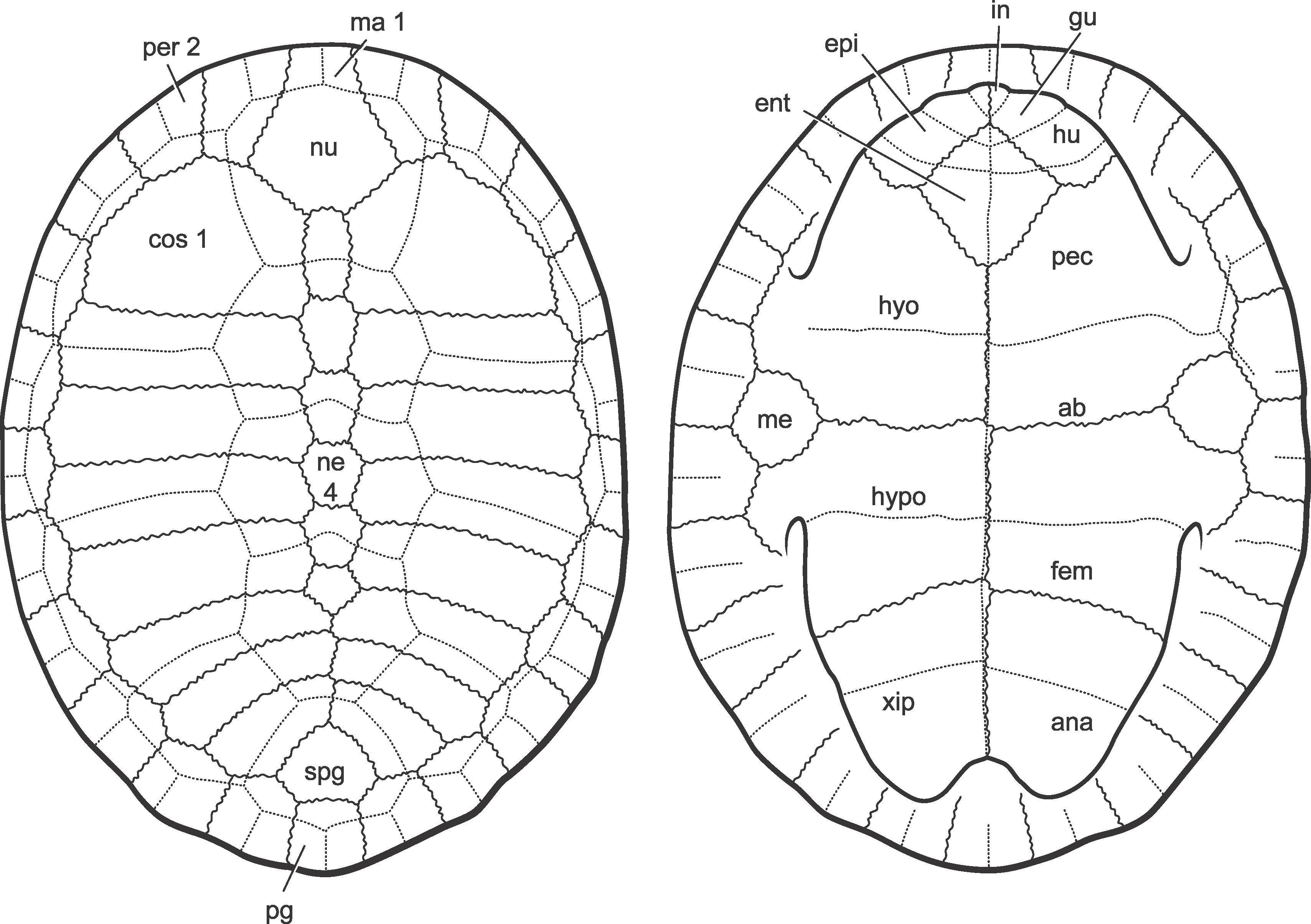
Fig. 47
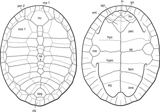
Neochelys fajumensis (Andrews, 1903). Restored shell based on literature, YPM, and AMNH material (see text). Left, dorsal; right ventral. [R. Wood, C. Wilson, del.]
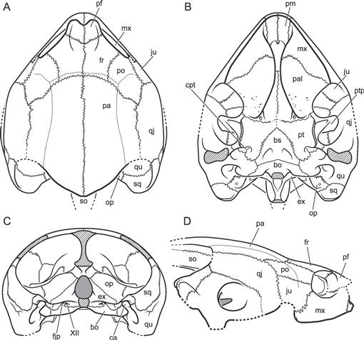
Fig. 48
Mogharemys blanckenhorni Dacqué (1912), n. gen. Partially restored skull based on BMNH R8440 and MB.R.2860 (cast of BMNH R8440). A, dorsal; B, ventral; C, lateral. [F. Ippolito, del.]
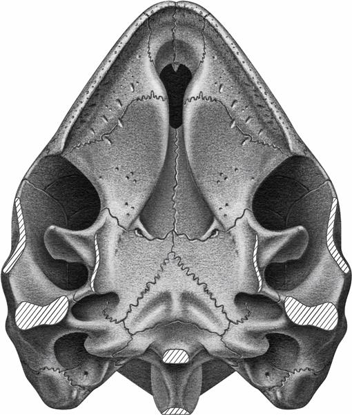
Fig. 49
Mogharemys blanckenhorni Dacqué (1912), n. gen. Partially restored ventral view based on BMNH R8440. [F. Ippolito, del.]
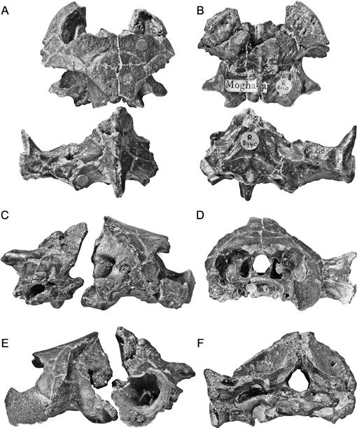
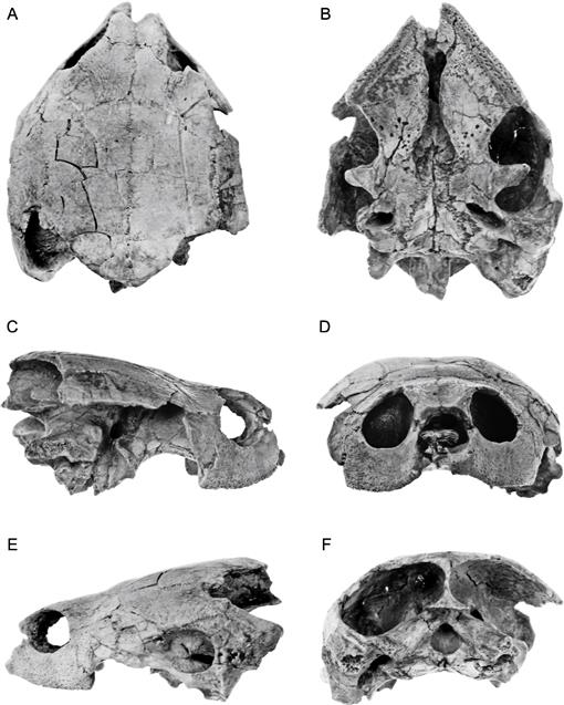
Fig. 50
Mogharemys blanckenhorni Dacqué (1912), n. gen. BMNH R8440. A, dorsal; B, ventral; C, right lateral; D, anterior; E, left lateral; F, posterior. [C. Facella, del.]
Fig. 51
Mogharemys blanckenhorni Dacqué (1912), n. gen. BMNH R8440. A, dorsal; B, ventral; C, right lateral; D, anterior; E, left lateral; F, posterior. [C. Facella, del.]
Fig. 52
Cordichelys antiqua (Andrews), n. gen. Partially restored skull based on YPM 7457. A, dorsal; B, ventral; C, occipital; D, lateral. [F. Ippolito, del.]
Fig. 53
Cordichelys antiqua (Andrews), n. gen. Partially restored ventral view of skull based on YPM 7457. [F. Ippolito, del.]
Fig. 54
Cordichelys antiqua (Andrews), n. gen. YPM 7457. A, dorsal; B, ventral; C, right lateral; D, anterior; E, left lateral; F, posterior. [F. Ippolito, del.]
Fig. 55
Cordichelys antiqua (Andrews), n. gen. YPM 7457. A, dorsal; B, ventral; C, right lateral; D, anterior; E, left lateral; F, posterior. [L. Starin, del.]
Fig. 56
Cordichelys antiqua (Andrews), n. gen. Shell of YPM 7457. A, dorsal; B, ventral. [E. Gaffney, del.]
Fig. 57
Cordichelys antiqua (Andrews), n. gen. Partially restored shell of YPM 7457. [C. Wilson, del.]
Fig. 58
Latentemys plowdeni, n. gen. et sp. Partially restored skull based on BMNH R.11998. A, dorsal; B, ventral; C, lateral. [C. Wilson, del.]
Fig. 59
Latentemys plowdeni, n. gen. et sp. Partially restored ventral view based on BMNH R.11998. [C. Wilson, del.]
Fig. 60
Latentemys plowdeni, n. gen. et sp. BMNH R.11998. A, dorsal; B, ventral; C, right lateral; D, anterior; E, left lateral; F, posterior. [G. Giardina, del.]
Fig. 61
Latentemys plowdeni, n. gen. et sp. BMNH R.11998. A, dorsal; B, ventral; C, right lateral; D, anterior; E, left lateral; F, posterior. [E. Heck, del.]
Fig. 62
Brontochelys gaffneyi (Wood), n. gen. Partially restored skull based on BMNH R.8570. A, dorsal; B, ventral; C, lateral. [E.E. Nixon, del.]
Fig. 63
Brontochelys gaffneyi (Wood), n. gen. Partially restored ventral view of skull based on BMNH R.8570. [E.E. Nixon, del.]
Fig. 64
Brontochelys gaffneyi (Wood), n. gen. BMNH R.8570. Skull. A, dorsal; B, ventral; C, right lateral; D, anterior; E, left lateral; F, posterior. [E.E. Nixon, del.]
Fig. 65
Brontochelys gaffneyi (Wood), n. gen. BMNH R.8570. Skull. A, dorsal; B, ventral; C, right lateral; D, anterior; E, left lateral; F, posterior. [E.E. Nixon, del.]
Fig. 66
Brontochelys gaffneyi (Wood), n. gen. BMNH R.8570. Medial views of internal cavum cranii. A, anterior moiety of right side; B, complete moiety of left side; C, restored sagittal view of left side. [J. Sharkey, del.]
Fig. 67
Lemurchelys diasphax, n. gen. et sp. Partially restored skull based on DPC 6425. A, dorsal; B, ventral; C, lateral. [I. Kayama, F. Ippolito, del.]
Fig. 68
Lemurchelys diasphax, n. gen. et sp. Partially restored ventral view based on DPC 6425. [I. Kayama, del.]
Fig. 69
Lemurchelys diasphax, n. gen. et sp. DPC 6425. Skull. A, dorsal; B, ventral; C, right lateral; D, anterior; E, left lateral; F, posterior. [E.E. Nixon, del.]
Fig. 70
Lemurchelys diasphax, n. gen. et sp. DPC 6425. Skull. A, dorsal; B, ventral; C, right lateral; D, anterior; E, left lateral; F, posterior. [F. Ippolito, del.]
Fig. 71
Shweboemys pilgrimi Swinton, 1939. Partially restored skull based on BMNH R.8432. A, dorsal; B, ventral; C, lateral. [F. Ippolito, del.]
Fig. 72
Shweboemys pilgrimi Swinton, 1939. Partially restored ventral view based on BMNH R.8432. [M. Rightmyer, del.]
Fig. 73
Shweboemys pilgrimi Swinton, 1939. BMNH R.8432. A, dorsal; B, ventral; C, right lateral; D, anterior; E, left lateral; F, posterior. [F. Ippolito, del.]
Fig. 74
Shweboemys pilgrimi Swinton, 1939. BMNH R.8432. A, dorsal; B, ventral; C, right lateral; D, anterior; E, left lateral; F, posterior. [F. Ippolito, del.]
Fig. 75
Stereogenys cromeri Andrews, 1901. Partially restored skull based on DPC 4120, BMNH R.3190, BMNH R.8442, BMNH R.3906, BMNH R.3189, BMNH R.3191. A, dorsal; B, ventral; C, lateral. [F. Ippolito, del.]
Fig. 76
Stereogenys cromeri Andrews, 1901. Partially restored ventral view based primarily on DPC 4120, with additions from BMNH R.3190, BMNH R.8442, and BMNH R.3191. A, dorsal; B, ventral; C, lateral. [F. Ippolito, del.]
Fig. 77
Stereogenys cromeri Andrews, 1901. DPC 4120. A, dorsal; B, ventral; C, right lateral; D, anterior; E, left lateral; F, posterior. [E. Heck, del.]
Fig. 78
Stereogenys cromeri Andrews, 1901. DPC 4120. A, dorsal; B, ventral; C, right lateral; D, anterior; E, left lateral; F, posterior. [M. Rightmyer, del.]
Fig. 79
Stereogenys cromeri Andrews, 1901. BMNH R.3191. Partial braincase and palate. A, dorsal; B, ventral; C, right lateral; D, anterior; E, left lateral. [E. Heck, del.]
Fig. 80
Stereogenys cromeri Andrews, 1901. BMNH R.3191. Partial braincase and palate. A, dorsal; B, ventral; C, right lateral; D, anterior; E, left lateral. [C. Wilson, del.]
Fig. 81
? Stereogenys cromeri Andrews, 1901. University of Michigan 161. Lower jaw. [N. Hennelly, del.]
Fig. 82
Stereogenys cromeri Andrews, 1901. DPC 4120. Endocast. [L. Starin, F. Ippolito, del.]
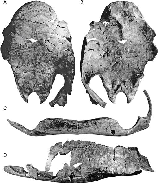
Fig. 83
Albertemys woodi, n. gen. et sp. AMNH 5088. Plastron and partial carapace. A, dorsal; B, ventral; C, anterior; D, left lateral. [R. Wood, del.]
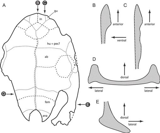
Fig. 84
Albertemys woodi, n. gen. et sp. AMNH 5088. Plastron and partial carapace. A, ventral view of plastron; B, C, D, E, cross sections at positions indicated. [R. Wood, C. Facella, del.]
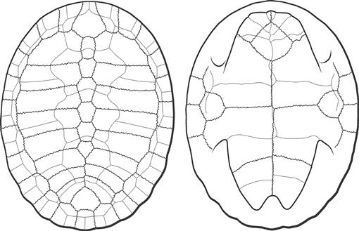
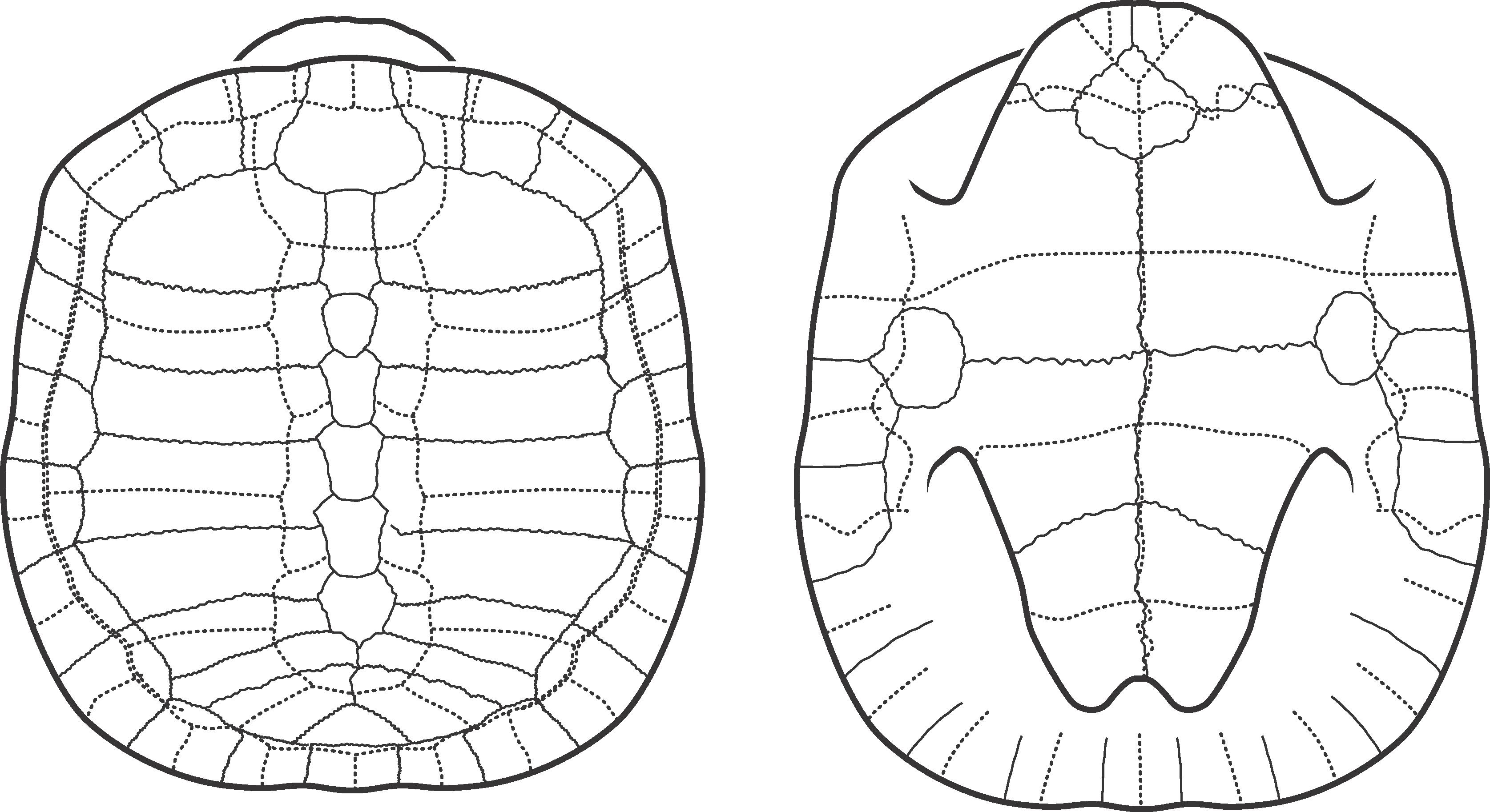
Fig. 85
“Stereogenys” libyca Andrews, 1903. Partially restored shell based on Andrews (1903: pl. 7, figs. A, B). [R. Wood, C. Wilson, del.]
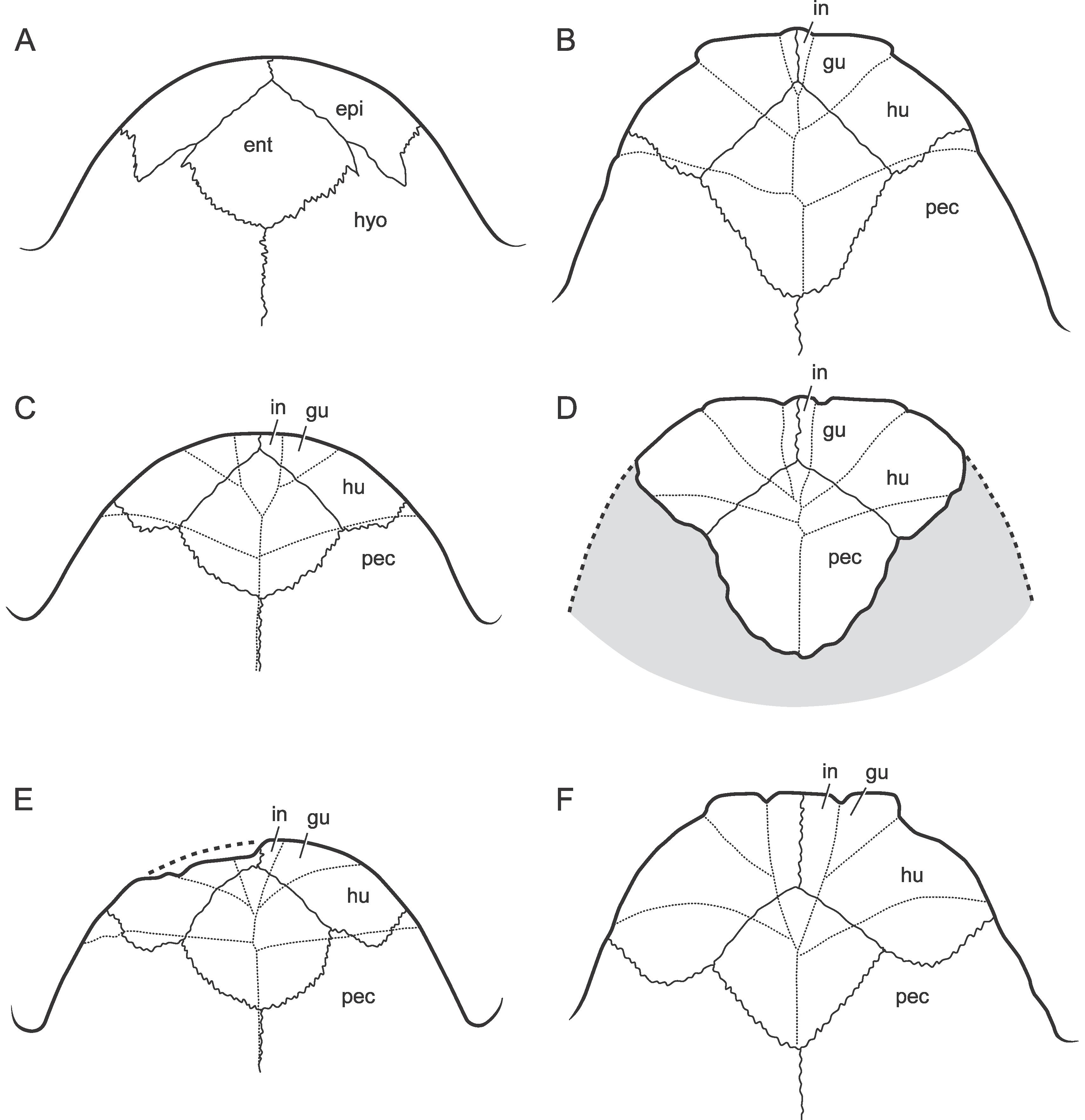
Fig. 86
Anterior lobes of Fayum podocnemidids. A, dorsal view of Cordichelys antiqua, n. gen. et sp., YPM 6205 (holotype); B, ventral view of Neochelys fajumensis, AMNH 5086; C, ventral view of Cordichelys antiqua, n. gen. et sp., YPM 6205 (holotype); D, ventral view of Neochelys fajumensis, AMNH 5093; E, ventral view of “Podocnemis stromeri” Reinach, 1903a, a synonym of C. antiqua; F, ventral view of “Stereogenys” podocnemoides Reinach, 1903b. Not to scale. [R. Wood, C. Wilson, del.]
Fig. 87
“Podocnemis” aegyptiaca Andrews, 1900. Restored shell, based on Andrews (1900: pl. 1) and Fourteau (1920: fig. 21). [R. Wood, C. Wilson, del.]
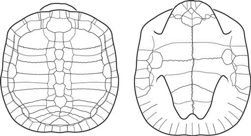
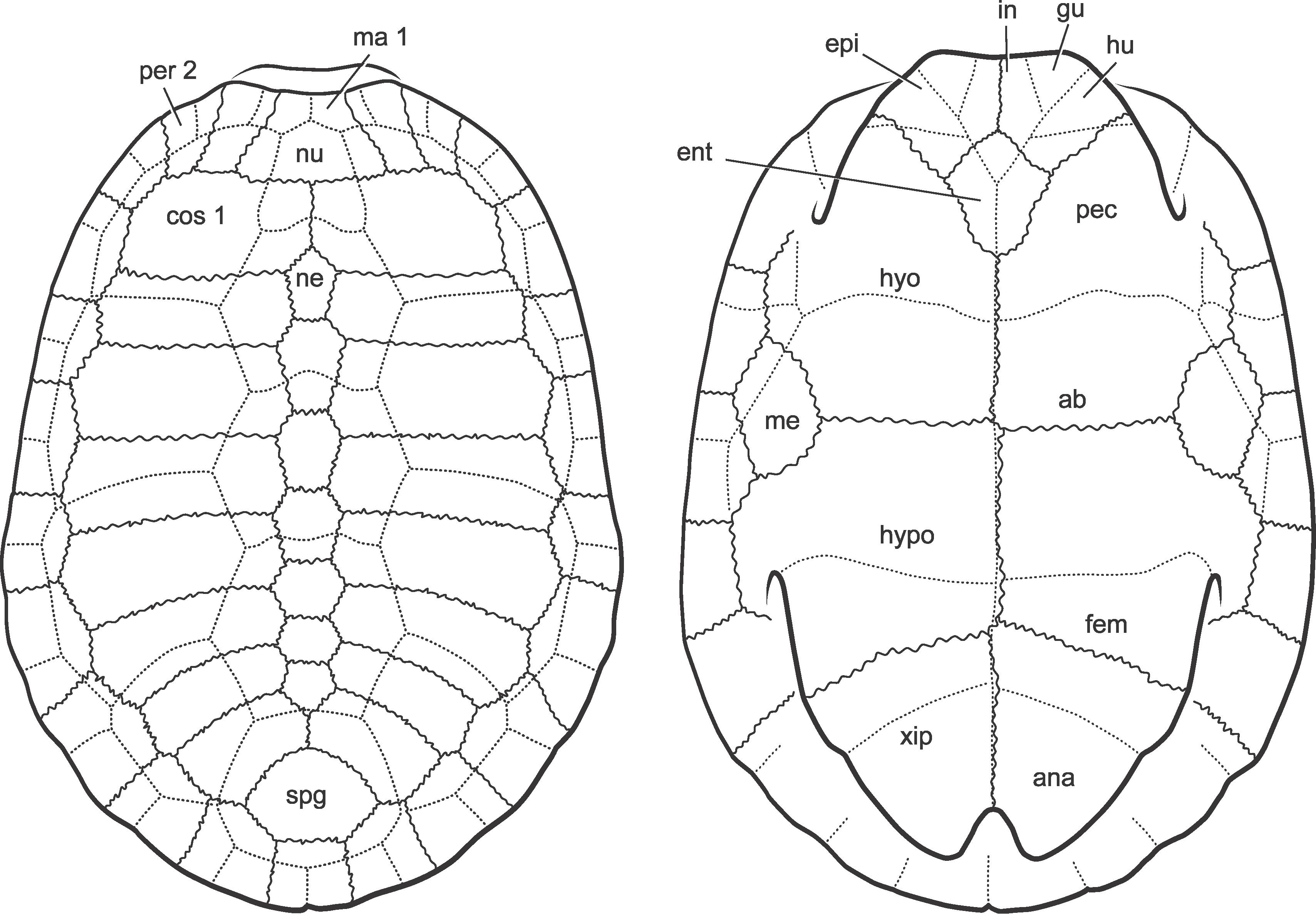
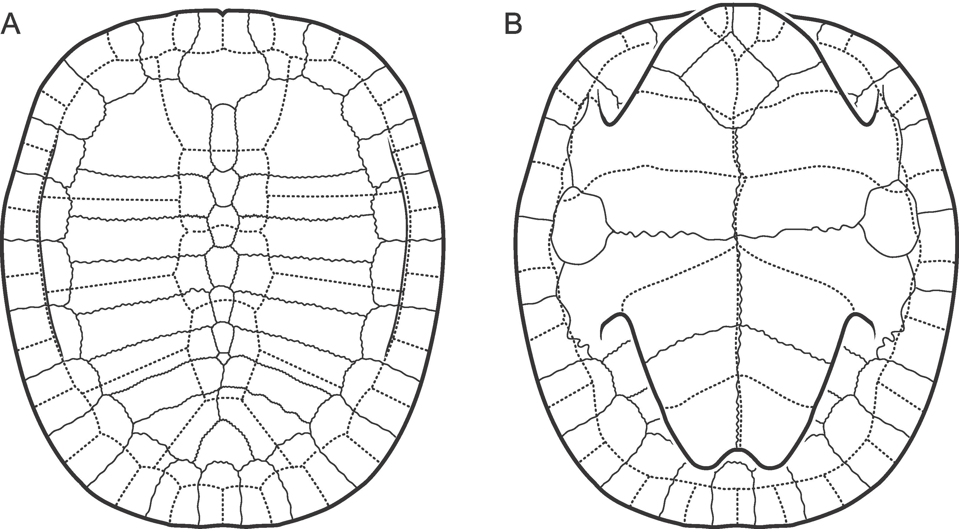
Fig. 88
Bauruemys elegans (Suárez, 1969a). Partially restored shell. [F. Ippolito, del.]
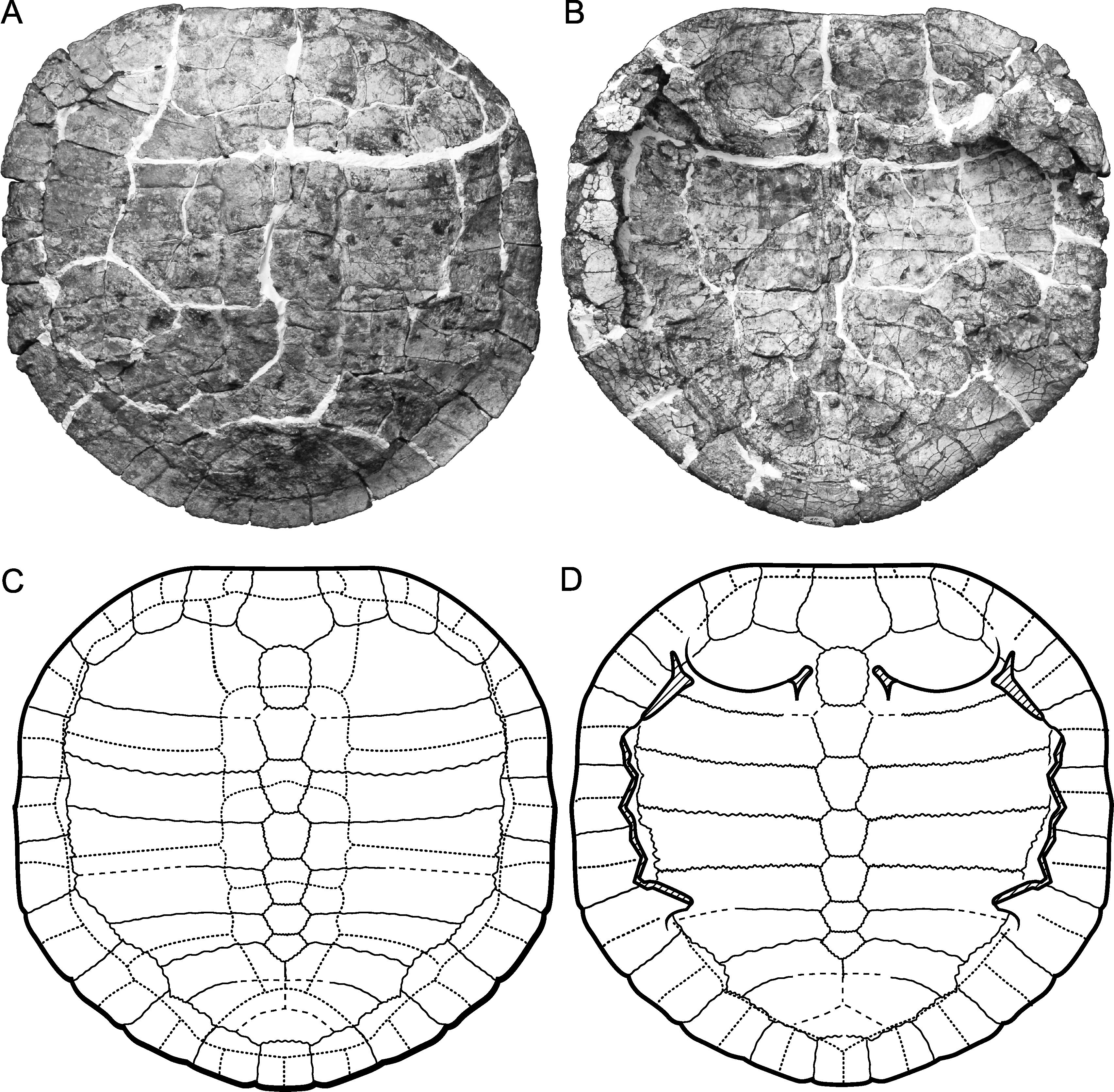
Fig. 89
Lapparentemys vilavilensis (Broin, 1971), n. gen. Shell. RM 20.5155. [F. Ippolito, del.]
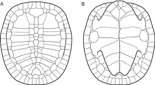
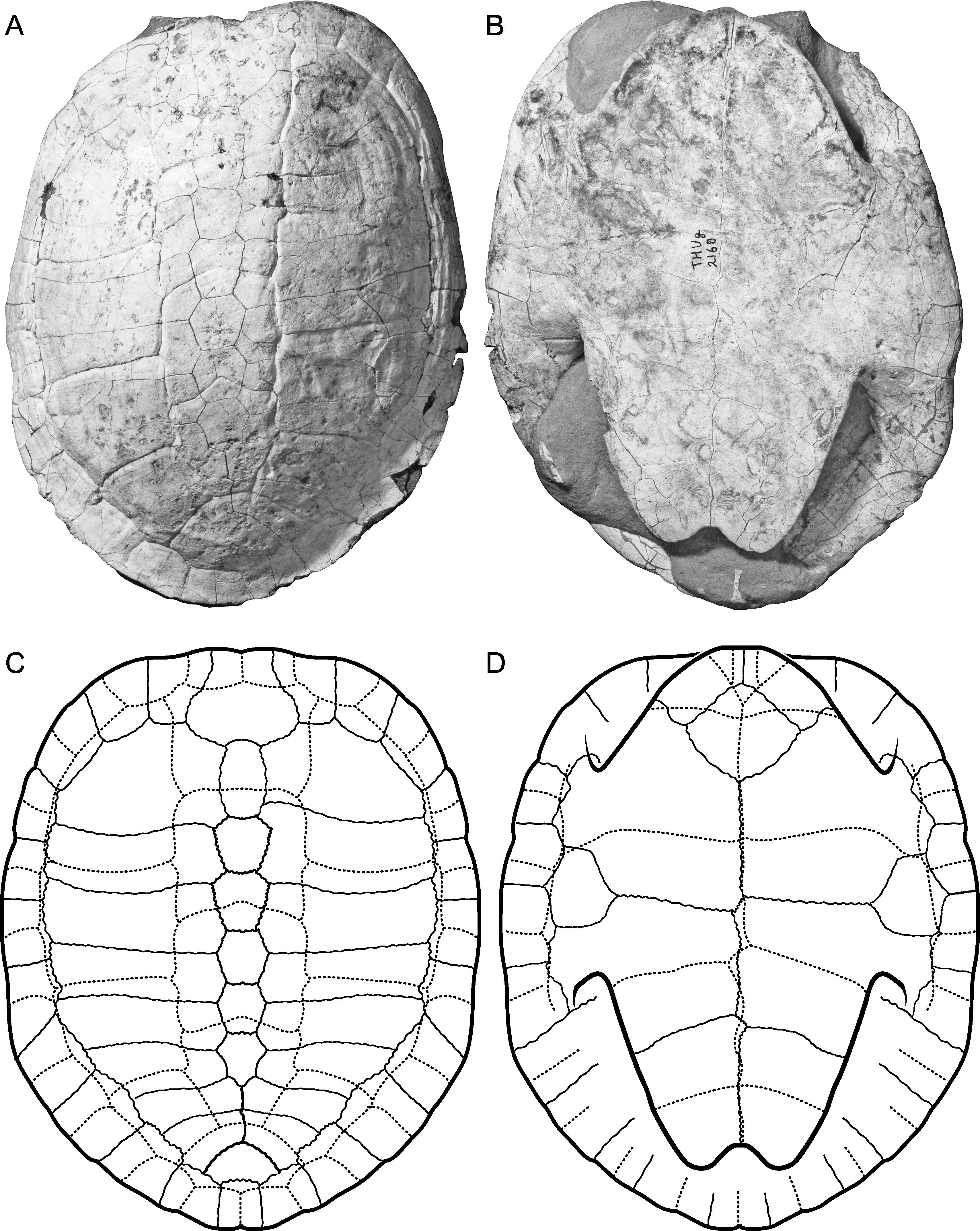
Fig. 90
Lapparentemys vilavilensis (Broin, 1971), n. gen. Shell. WUS 2160. [F. Ippolito, del.]
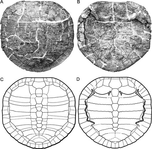
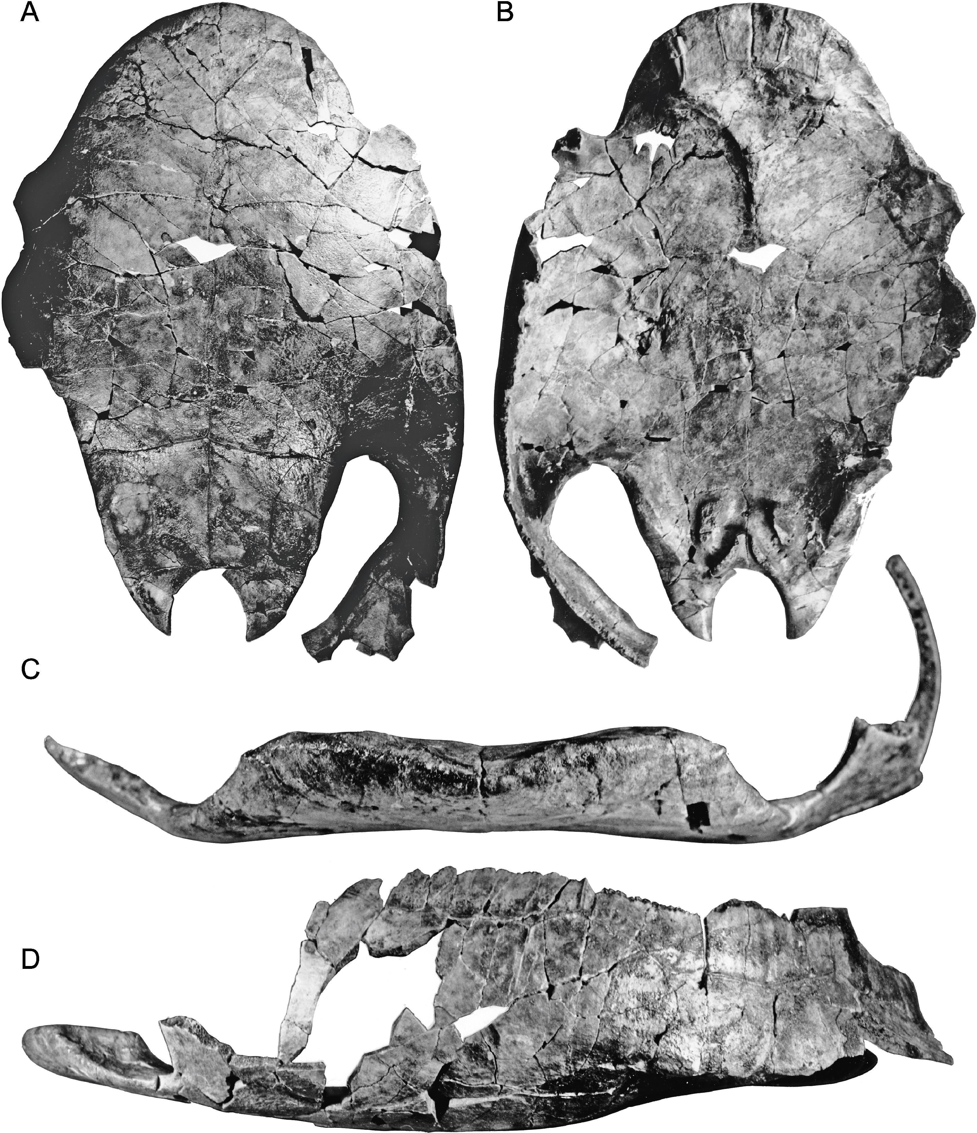
Fig. 91
Shell attributed to unnamed taxon Peirópolis A. A, Carapace speculatively restored from disarticulated parts (see text); B, plastron, DGM MCT 1499-R, a complete shell, with unprepared carapace. [F. Ippolito, del.]
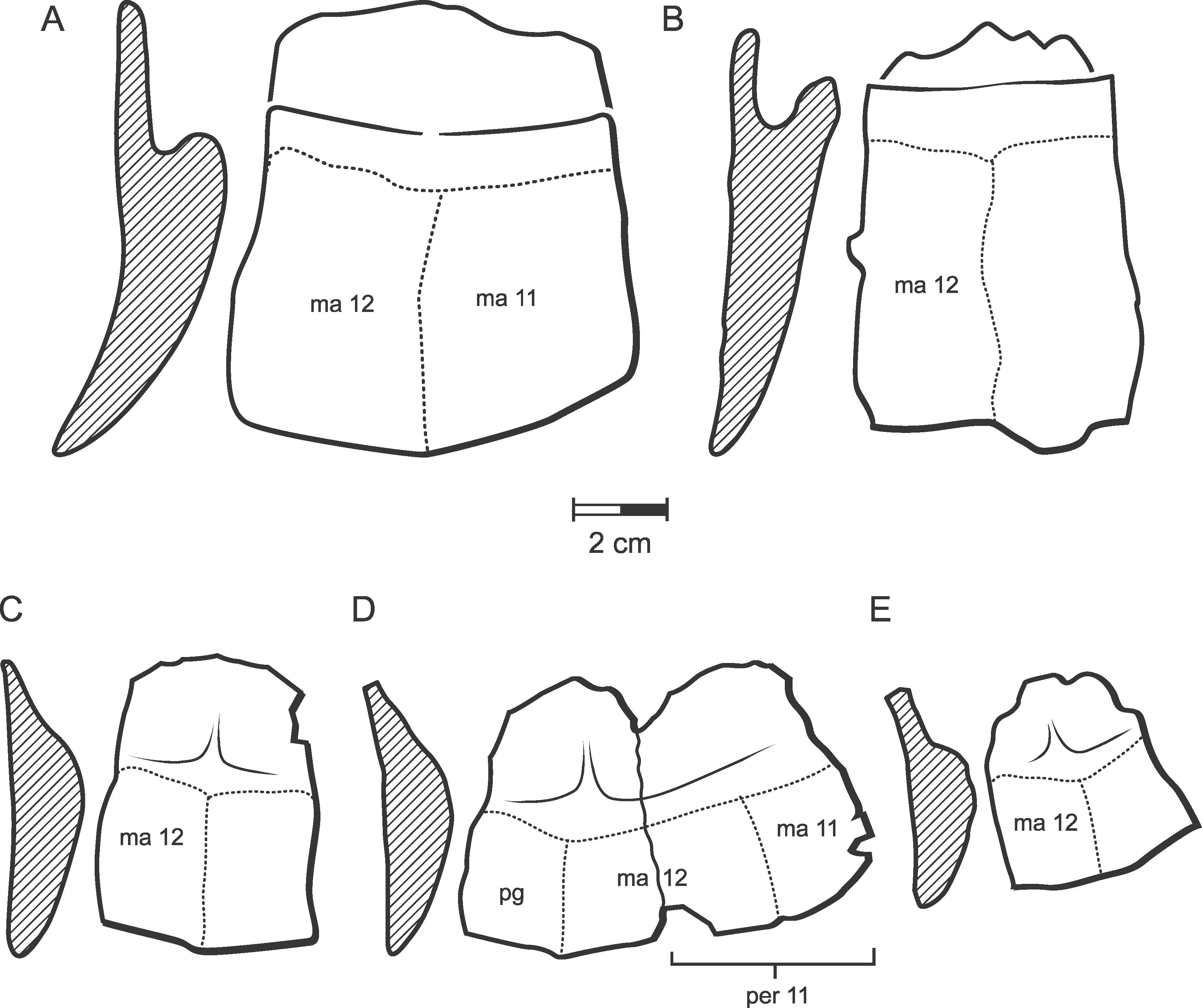
Fig. 92
Pygal and adjacent peripheral bones attributed to unnamed taxa Peirópolis A (upper row) and Peirópolis B (lower row) in ventral view, side views on left. All are DNPM DGM uncataloged (see text entries) from Caiera Quarry, near Uberaba, Minas Gerais State, Brazil. Serra da Galga Member, Marília Fm., Maastrichtian. See text entries for Peirópolis A and Peirópolis B (also Peiropemys mezzalirai, n. gen. et sp.) for further information. A, Peirópolis A pygal; B, Peirópolis A peripheral 11; C, Peirópolis B pygal; D, Peirópolis B pygal with articulated left peripheral 11 attached E, Peirópolis B pygal. [F. Ippolito, del.]
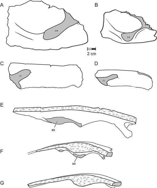
Fig. 93
Costal bones attributed to unnamed taxa Peirópolis A and Peirópolis B (see text entries for these taxa), showing axillary and inguinal buttress attachment sites. All are DNPM DGM uncataloged from Caiera Quarry, near Uberaba, Minas Gerais State, Brazil. Serra da Galga Member, Marília Fm., Maastrichtian. See text entries for Peirópolis A and Peirópolis B (also Peiropemys mezzalirai, n. gen. et sp.) for further information. A, first left costal in ventral view, Peirópolis A; B, first left costal in ventral view, Peirópolis B; C, fifth right costal in ventral view, Peirópolis A; D, fifth right costal in ventral view, Peirópolis B; E, Peirópolis A, first left costal in posterior view, midline to right; F, Peirópolis B, first left costal inposterior view, midline to right; G, Peirópolis B, second right costal in anterior view to show swelling of costals one and two contact behind axillary buttress. [F. Ippolito, del.]
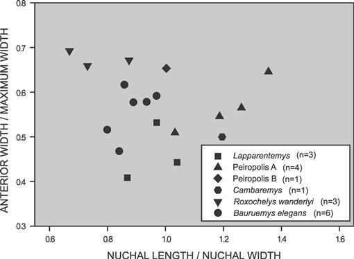
Fig. 94
Plot of nuchal shape in some South American Cretaceous and Early Tertiary podocnemidids. [P. Meylan, F. Ippolito, del.]
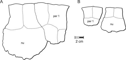
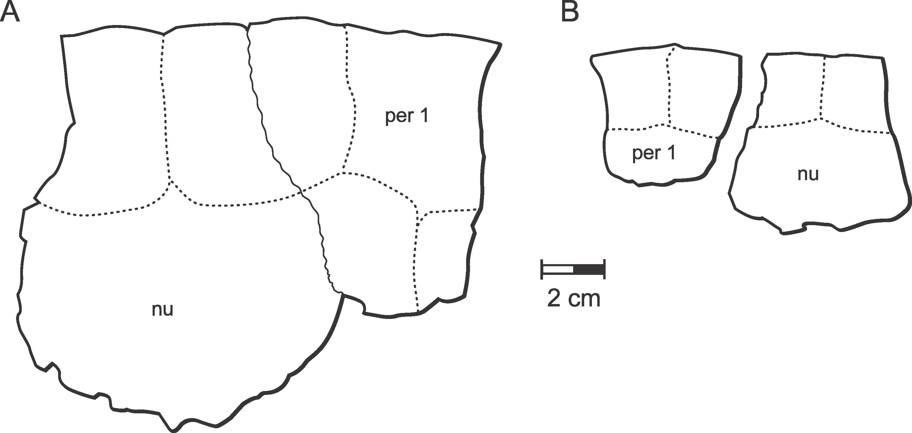
Fig. 95
Nuchals attributed to unnamed taxa, Peirópolis A and Peirópolis B (see text entries for further discussion). All are DNPM DGM uncataloged from Caiera Quarry, near Uberaba, Minas Gerais State, Brazil. Serra da Galga Member, Marília Fm., Maastrichtian. A, nuchal and first right peripheral of Peirópolis A; B, nuchal and first left peripheral of Peirópolis B. [F. Ippolito, del.]
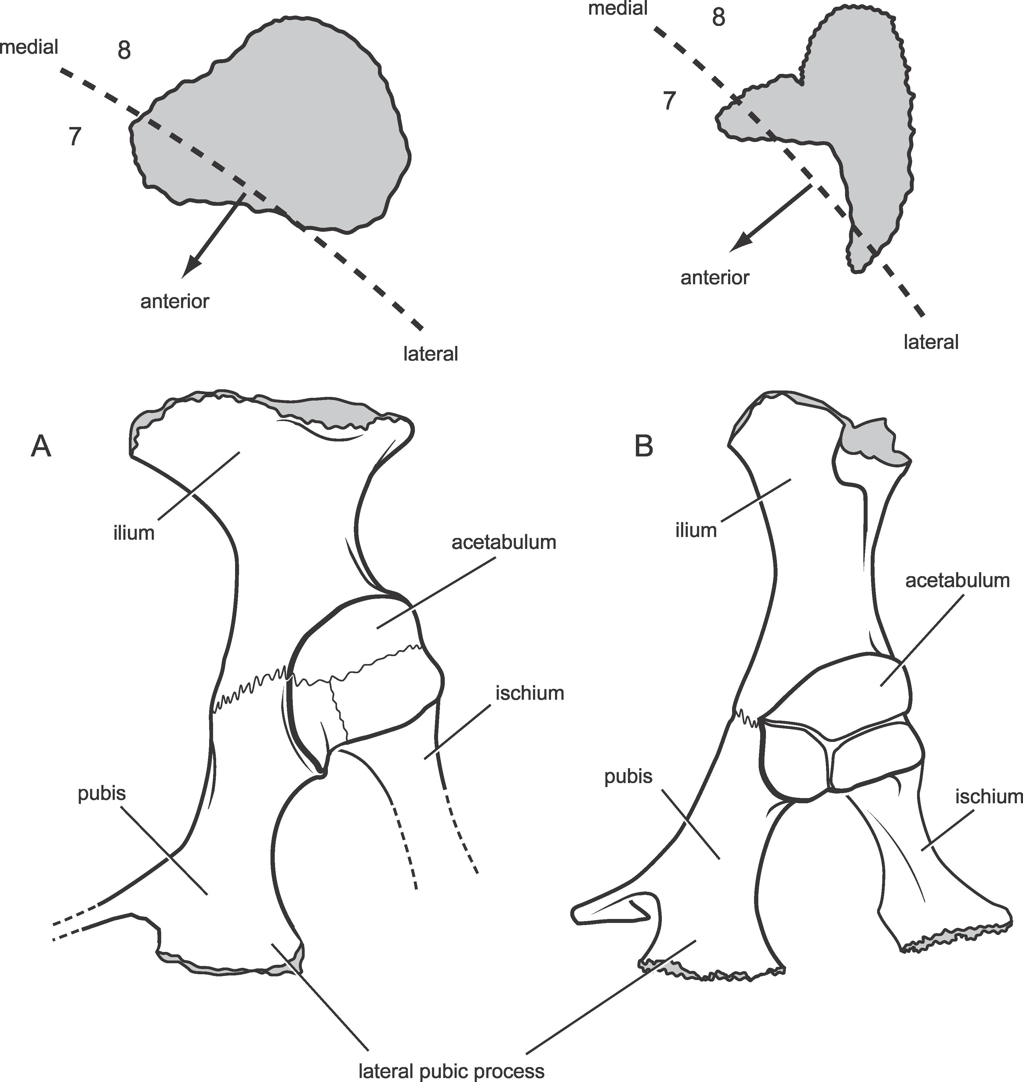
Fig. 96
Pelves and iliac attachment sites of podocnemidids, left pelvis in lateral view with iliac sutural area indicated above showing position of costals seven and eight. A, Peirópolis A, DNPM DGM uncataloged from Caiera Quarry, 1974, near Uberaba, Minas Gerais State, Brazil. Serra da Galga Member, Marília Fm., Maastrichtian. B, Podocnemis expansa AMNH 62947, Recent. [F. Ippolito, del.]
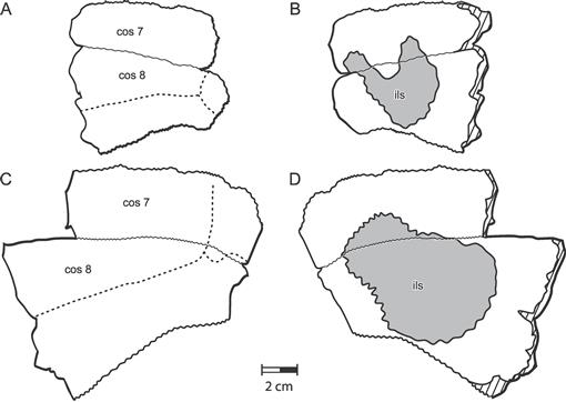
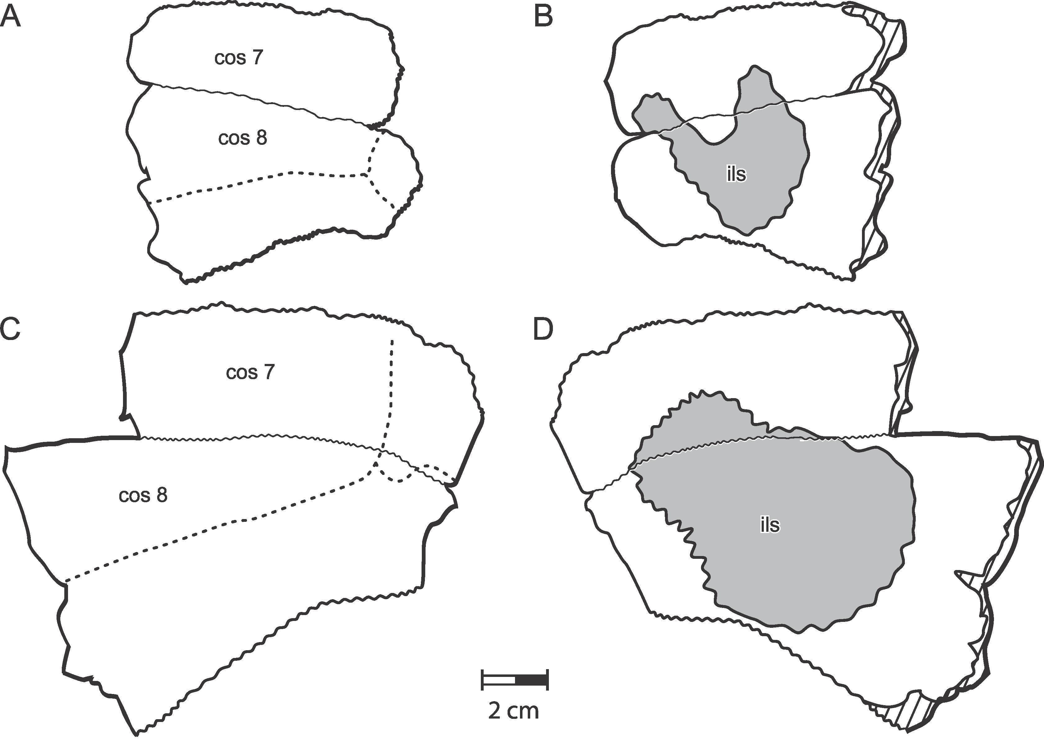
Fig. 97
Comparison of left costals seven and eight attributed to unnamed taxa Peirópolis A and Peirópolis B (see text entries for these taxa). All are DNPM DGM uncataloged from Caiera Quarry, near Uberaba, Minas Gerais State, Brazil. Serra da Galga Member, Marília Fm., Maastrichtian. Anterior toward top of page. A, C, dorsal views, medial to right; B, D, ventral views, medial to left. A and B are hypothesized as Peirópolis B; C and D are hypothesized as Peirópolis A. [F. Ippolito, del.]
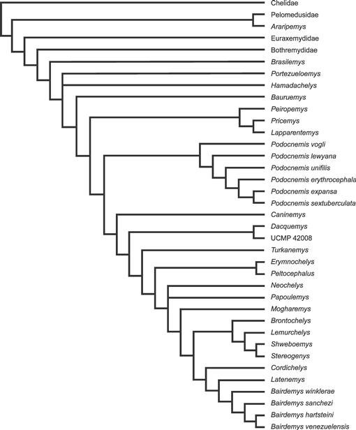
Fig. 98
Consensus cladogram of nine equally parsimonious cladograms of 173 steps resulting from a PAUP* analysis of 74 characters (69 informative) and 37 taxa. All characters unweighted and unordered, character matrix in appendix 1, character list in appendix 2. [F. Ippolito, del.]
(See Gaffney et al., 2006: 685, fig. 315 for positions of measurements. D1 is left orbital width; D2 is right orbital width; J1 is height of left orbit.) All measurement in mm.


