Open Access
How to translate text using browser tools
28 July 2017 Morphology and ultrastructure of Tetranychusturkestani Ugarov & Nikolskii (Acari: Tetranychidae)
Jue-Ying-Qi Jiang, Yan-Nan Zhang, Dan-Dan Guo, Jian-Ping Zhang, Jing Chen
Author Affiliations +
FIGURE 1.
All stages of Tetranychus turkestani (LM). A, Egg; B, Larva; C, Protonymph; D, Deutonymph; E, Female adult; F, Male adult.

FIGURE 2.
Tetranychus turkestani (adult female, LM). A, Dorsal view; B, Ventral view; C, Stylet (St) in ventral view; D, Peritreme (Pe) and stylophore (Sp) in dorsal view.

FIGURE 3.
Tetranychus turkestani (adult female, SEM). A, Gnathosoma in lateral view, showing eye (Eye), palpfemoral seta (d), palpus (Pa), and stylophore (Sp); B, Pedipalp in lateral view, showing trochanter (Tr), femur (Fe), genu (Ge), tibia (Ti) and tarsus (Ta); C, Gnathosoma in ventral view, showing rostrum (Ro), subcapitular setae (m) and subcapitulum (Sub); D, Palptarsus in ventral view, showing palpotibial claw (CI), palpotarsal eupathidial spinneret (suζ), eupathidium (ul′ζ and ul′ζ), setae (a, b and c), solenidion (ω); E, Opisthosoma in ventral view, showing aggenital setae (ag), anal opening (AO), genital flap (GF), genital opening (GO), genital setae (g1, and g2), para-anals (h3), post-anals (h2) and pseudanal setae (ps1 and ps2); F, Genital region.

FIGURE 4.
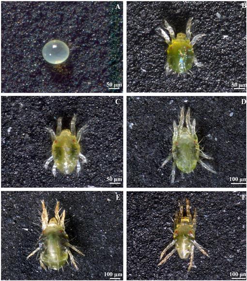
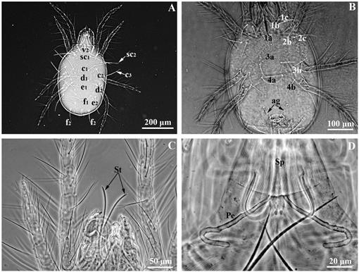
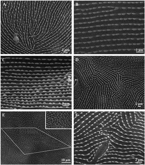
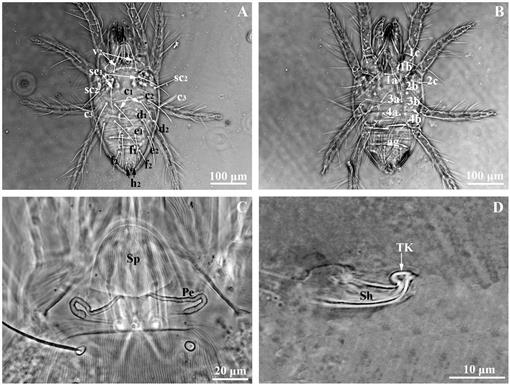
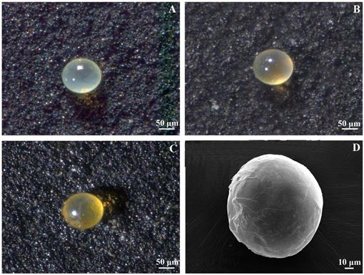
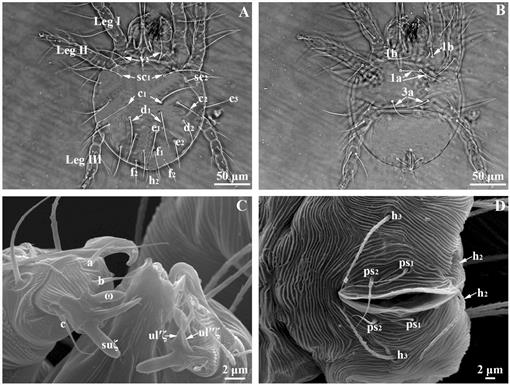
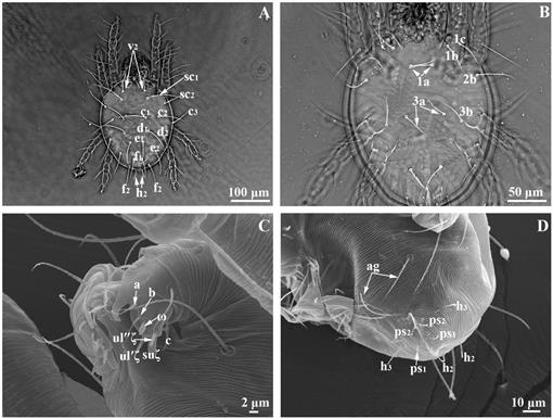
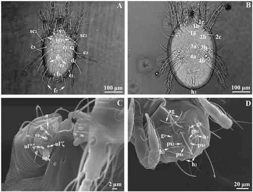
Tetranychus turkestani (adult female, SEM). A, The striae and integumentary lobes between the setae v2, and sc1; B, C The striae and integumentary lobes between the setae c1, d1 and e1; D, The striae and integumentary lobes between the setae e1; E, The striae and integumentary lobes between the setae e1 and f1; F, lyrifissure (Ly).

FIGURE 5.
Tetranychus turkestani (adult female, SEM). A, Leg I in ventral view, showing coxa (Cx), trochanter (Tr), femur (Fe), genu (Ge), tibia (Ti), tarsus (Ta) and pretarsus (Pr); B, Tibia (Ti) and Tarsus (Ta) I in lateral view; C, Duplex setae (ω′,ft′ and ω″, ft″); D, Tarsus and pretarsus I in lateral view, showing empodium (Em), prorals (p′ζ), tenent hairs (TH), tectal setae (tc′) and primiventrals (pv).

FIGURE 6.
Tetranychus turkestani (adult male, LM). A, Dorsal view; B, Ventral view; C, Peritreme (Pe) and stylophore (Sp) in dorsal view; D, Aedeagus in lateral view, showing shaft (Sh) and terminal knob (TK).

FIGURE 7.
Tetranychus turkestani (adult male, SEM). A, B, Gnathosoma in dorsal view, showing palpi (Pa), rostral gutter (RG), stylophore (Sp), stylet (St), palpfemoral seta (d), trochanter (Tr), femur (Fe), genu (Ge), tibia (Ti) and tarsus (Ta); C, Gnathosoma in ventral view, showing palpotarsal eupathidial spinneret (suζ), eupathidium (ul′ζ and ul″ζ), rostrum (Ro), setae (a, b and c), solenidion (ω), subcapitular setae (m) and subcapitulum (Sub); D, Opisthosoma in ventral view, showing genital setae (g1 and g2), para-anals (h3) and pseudanal setae (ps1 and ps2).

FIGURE 8.
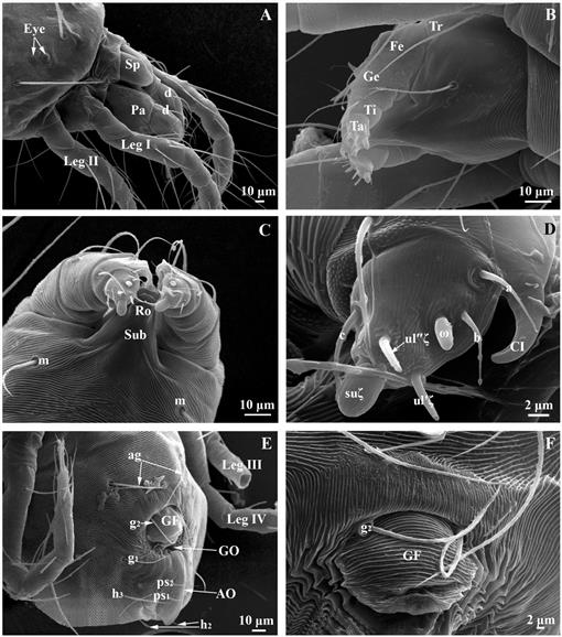
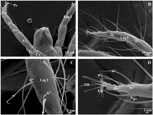
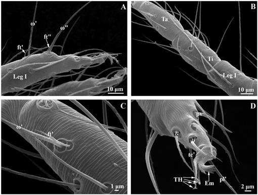
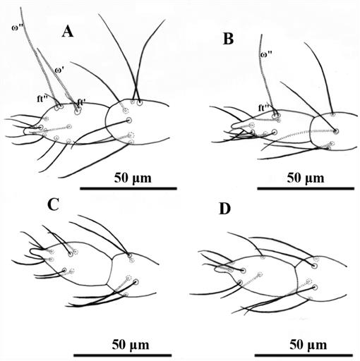
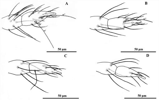
Tetranychus turkestani (adult male, SEM). A, Tarsus and pretarsus I in lateral view; B, Tibia (Ti) and Tarsus (Ta) I in lateral view; C, Duplex setae (ω′ and ƒt′); D, Tarsus and pretarsus I in lateral view, showing empodium (Em), primilaterals (pl′ and pl″), primiventrals (pv), tenent hairs (TH) and tectal setae (tc′).

FIGURE 9.
Tetranychus turkestani (egg, LM, SEM). A, 24 hr; B, 48 hr; C, 72 hr; D, 48 hr.

FIGURE 10.
Tetranychus turkestani (larva, LM, SEM). A, Dorsal view; B, Ventral view; C, Palptarsus in ventral view, showing eupathidial spinneret (suξ), eupathidium (ul′ξ and ul″ξ), setae (a, b and c), and solenidion (ω); D, Opisthosoma in ventral view, showing para-anals (h3). post-anals (h2) and pseudanal setae (ps1 and ps2).

FIGURE 11.
Tetranychus turkestani (larva). A, Leg I; B, Leg II; C, Leg III.

FIGURE 12.
Tetranychiis turkestani (protonymph, LM, SEM). A, Dorsal view; B, Ventral view; C, Palptarsus in ventral view, showing eupathidial spinneret (suξ), eupathidium (ul′ξ and ul″ξ), setae (a, b and c) and solenidion (ω); D, Opisthosoma in ventral view, showing aggenital setae (ag), para-anals (h3), post-anals (h2) and pseudanal setae (ps1 and ps2).

FIGURE 13.
Tetranychiis turkestani (protonymph). A, Tibia and tarsus I; B, Tibia and tarsus II; C, Tibia and tarsus III; D, Tibia and tarsus IV.

FIGURE 14.
Tetranychus turkestani (deutonymph, LM, SEM). A, Dorsal view; B, Ventral view; C, Palptarsus in ventral view, showing eupathidial spinneret (suξ), eupathidium (ul′ξ and ul″ξ), setae (a, b and c) and solenidion (ω); D, Opisthosoma in ventral view, showing aggenital setae (ag), genital setae (g1). para-anals (h3), post-anals (h2) and pseudanal setae (ps1 and ps2).

FIGURE 15.
Tetranychus turkestani (deutonymph). A, Tibia and tarsus I; B, Tibia and tarsus II; C, Tibia and tarsus III; D, Tibia and tarsus IV.

TABLE 1.
The length of dorsal setae of T. turkestani at different development stages (n=10).

KEYWORDS
adult
deutonymph
egg
larva
protonymph
spider mite
Back to Top