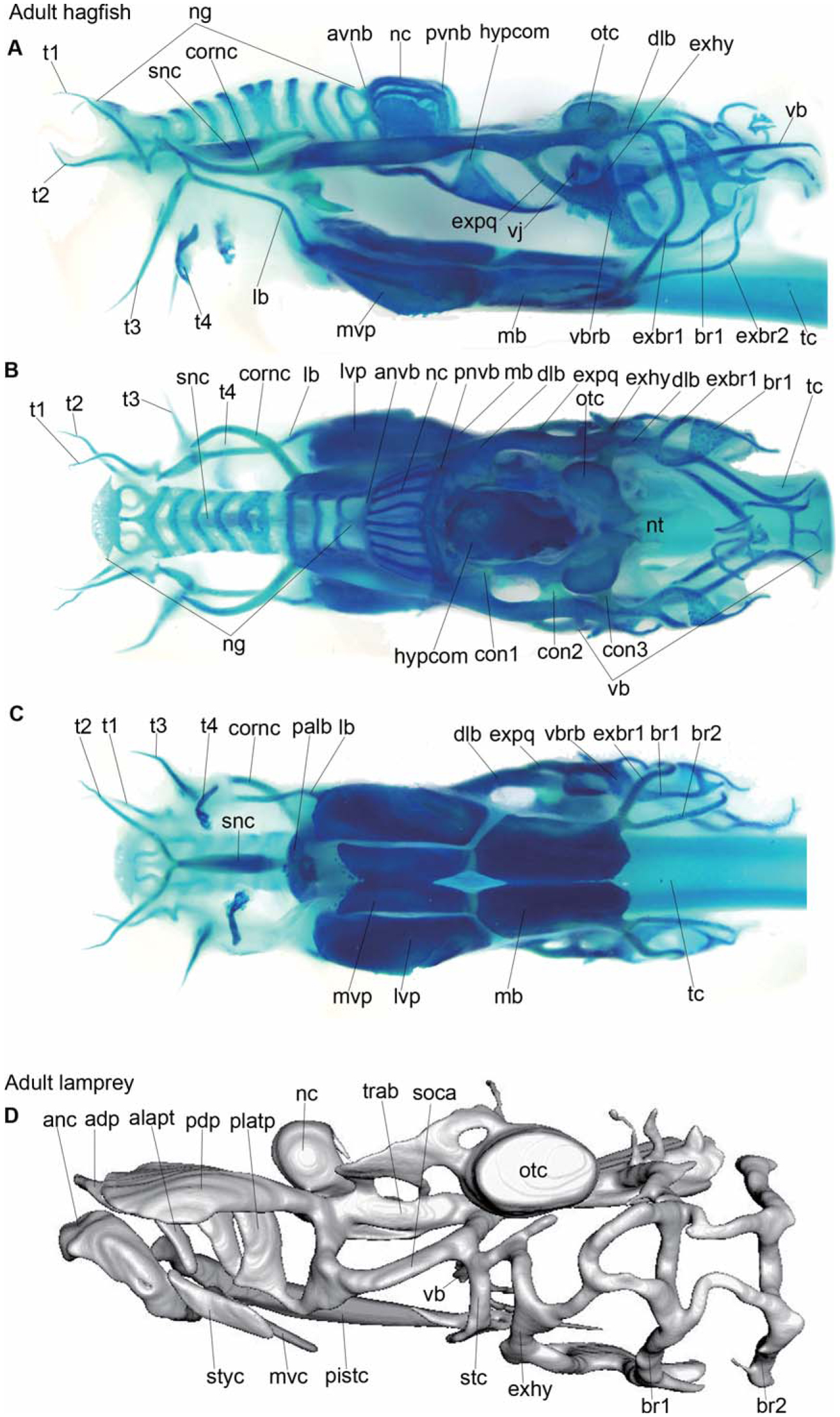
Fig. 1.
Adult chondrocrania of cyclostomes. Lateral (A), dorsal (B), and ventral (C) views of an Alcian-blue-stained whole-mount chondrocranium of Eptatretus burgeri, and 3D reconstructed model of the cranium in an adult lamprey, Lethenteron reissneri (D). See the list in the text for abbreviations.
Fig. 2.
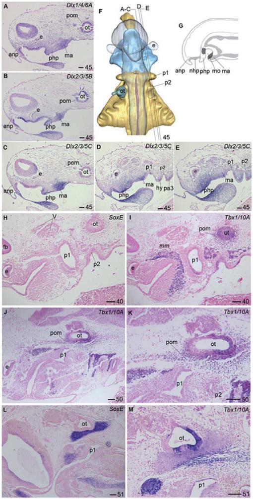
Embryonic expression of genes for craniofacial structure in Eptatretus burgeri. Histologic sections of E. burgeri at stages 45 (A–E), 40 (H, I), 50 (J, K), and 51 (L, M), hybridized with Dlx1/4/6A (A), Dlx2/3/5B (B), Dlx2/3/5C (C–E), Tbx1/10A (l–K, M), or SoxE (H, L) riboprobes. Also dorsal view of reconstruction (F) and schematic diagram of Dlx genes expression in the mid-sagittal region (G) of a stage 45 embryo (dark gray). At stage 40 (I), Tbx1/10A, the mesodermal neurocranial marker gene, is expressed in the pharyngeal arch muscle anlage as well as in the mesenchyme surrounding the otocyst, representing the otic capsule anlage. By stage 50 (J, K), the mandibular arch muscle anlage has differentiated into tentacular and lingual muscle primordia, and the periotic prochondrogenic mesenchyme has grown rostrally to form the common anlage for trabeculae and the dorsal longitudinal bar. (L) SoxE expression at stage 51 depicts a rostrally growing longitudinal prochondrogenic anlage, continuous with the otic capsule. Abbreviations: anp, anterior nasal process; e, eye; mm, mandibular mesoderm; ot, otocyst; p1, 2, pharyngeal pouches 1, 2; php, posthypophyseal process; pom, periotic mesenchyme. See the list in the text for other abbreviations. Bars = 100 µm.
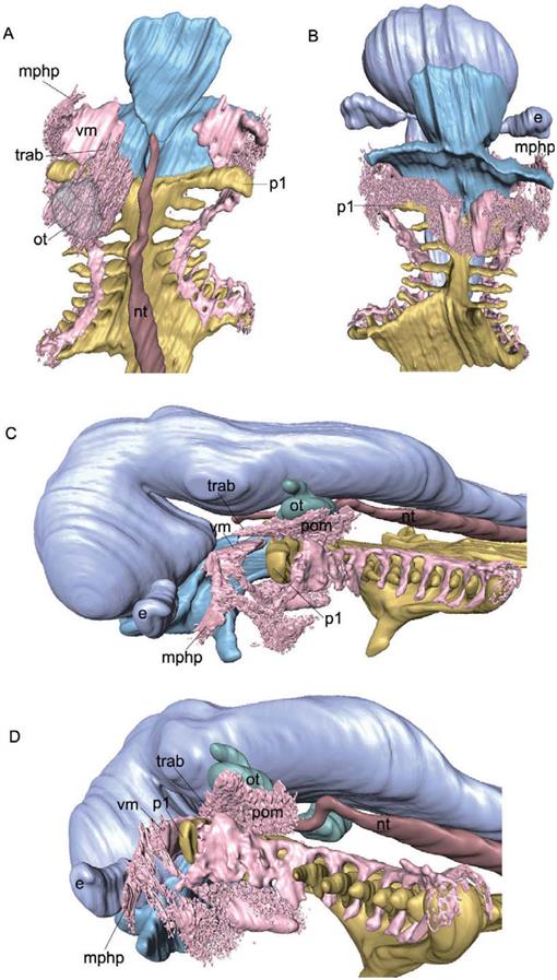
Fig. 3.
Mesenchymal cranial primordium of a stage 50 Eptatretus burgeri embryo. 3D reconstructions were based on sectioned specimens hybridized with Tbx1/10A (A–D) riboprobes. (A) Dorsal view. (B) Ventral view. (C) Left lateral view. (D) Oblique posterior view. The ectodermal oronasohypophyseal cavity is colored light blue, the pharyngeal endoderm is colored yellow, and the Tbx1/10A positive-mesoderm colored pink, mphp, muscle primordia in the php; vm, velum muscle; nt, notocord; trab, trabecula. See the list in the text for other abbreviations.
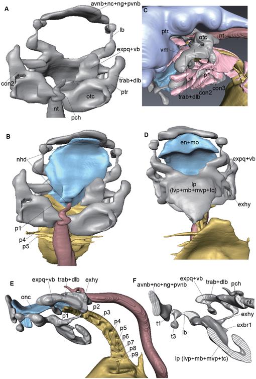
Fig. 4.
Prochondrogenic cranial primordium of a stage 51 Eptatretus burgeri embryo. 3D-reconstructions were based on SoxE and Tbx1/10A expression. (A, F) Reconstructions of the cranial primordium. (B–E) Reconstructions with epithelial structures (ectodermal oronasohypophyseal cavity, light blue; pharyngeal endodermal lining, yellow; Tbx1/10A-positive head muscle primordium, pink). (A, B) Dorsal views. (D) Ventral views. (E, F) Left lateral views. lp, lingual plate; otc, otic capsule; t1 and t3, common anlage for tentacular cartilages I and III; trab + dlb, common anlage for trabeculae and the dorsal longitudinal bar; vb, velar bar. See the list in the text for other abbreviations.
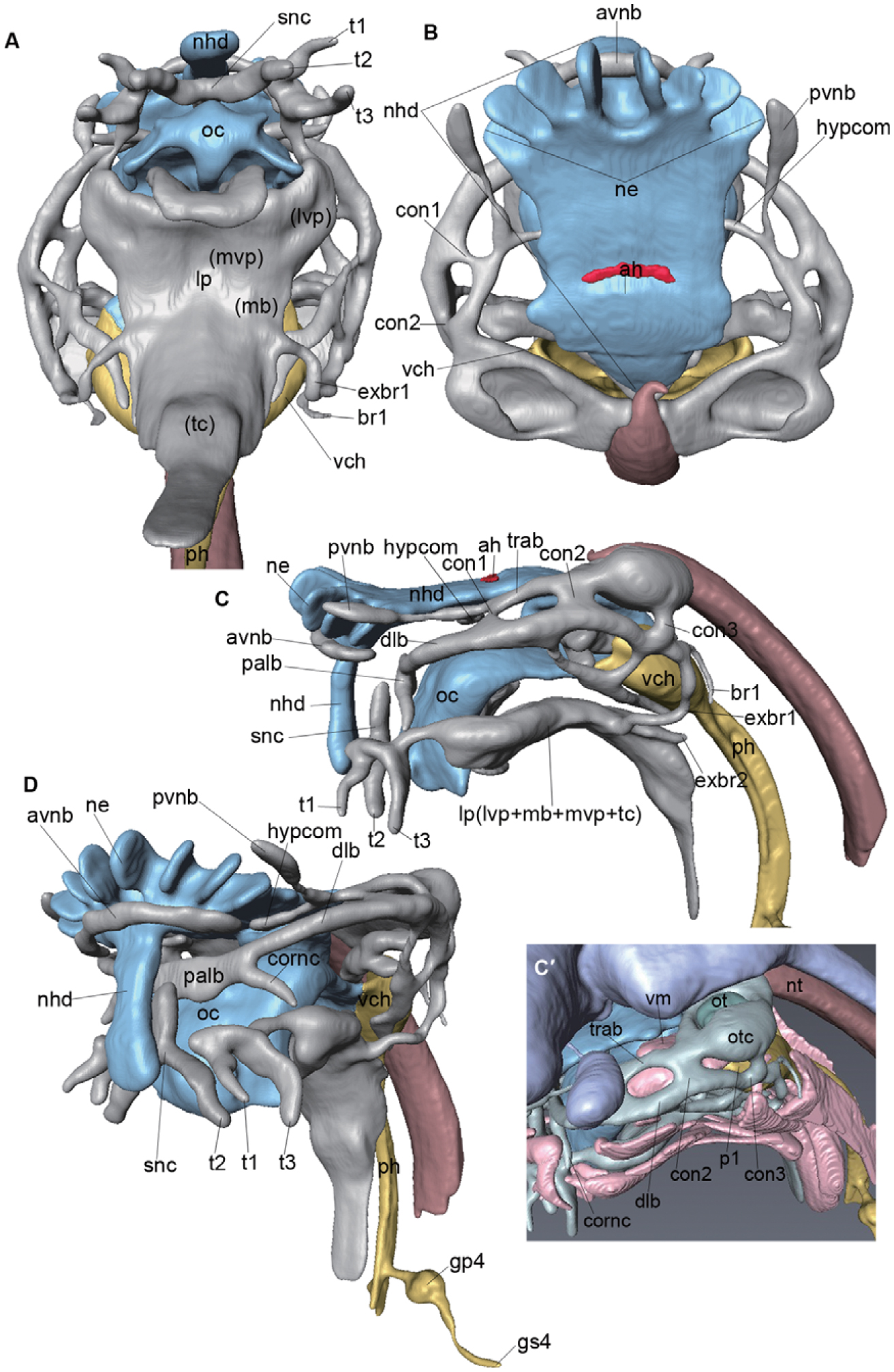
Fig. 5.
Chondrocranium of a stage 53 Eptatretus burgeri embryo. Ventral (A, C), dorsal (B), and left lateral (D) views of a 3D-reconstructed model. Lingual cartilage is removed in (C). See the list in the text for abbreviations.
Fig. 6.
The same chondrocranium as shown in Fig. 5, reconstructed with endodermal (yellow) and ectodermal (light blue) linings associated with the cranium. The Tbx1/10A-positive head muscle primordium is coloured pink.Ventral (A), dorsal (B), lateral (C, C′), and oblique anterior (D) views of a 3D-reconstructed model. Note in the lateral view (C) that most of trabeculae (trab) lies below the nasohypophyseal duct, or within the oronasohypophyseal septum (space between nasohypophyseal duct and oral cavity). The lingual plate lies below the oral cavity, or within the ventral part of the mandibular arch. See the list in the text for abbreviations.
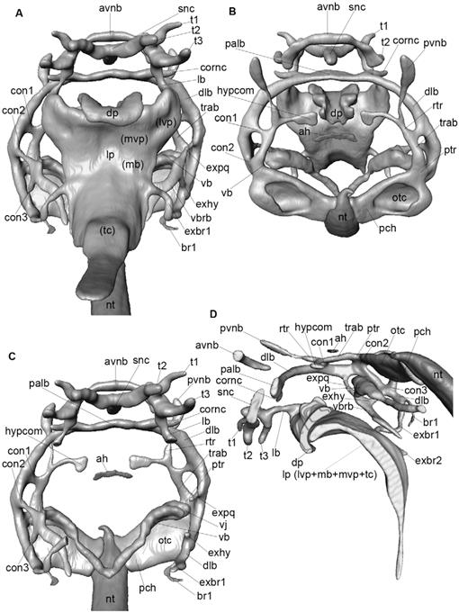
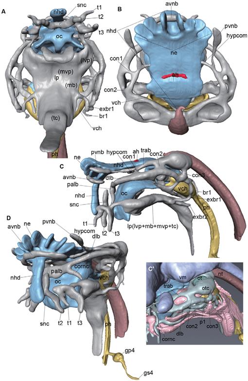
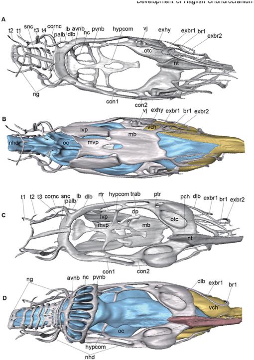
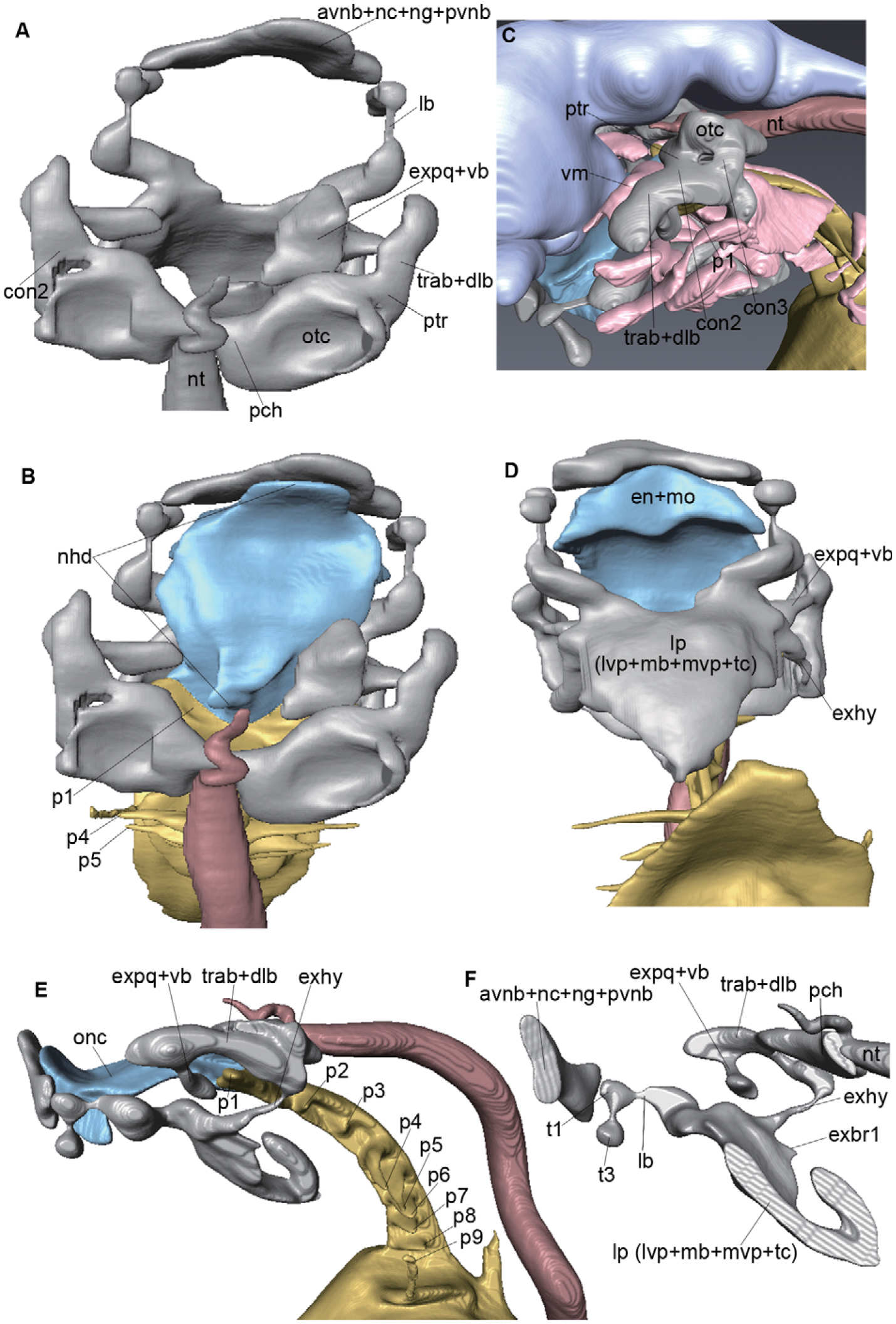
Fig. 7.
Chondrocranium of a stage 60 Eptatretus burgeri embryo. (B, C, F, G) Reconstruction of the chondrocranium. (A, D, E, H) Reconstruction with epithelial structures (ectodermal oronasohypophyseal cavity, light blue; pharyngeal endodermal lining, yellow). (A, B) Dorsal views. (C, D) Ventral views. (E, F) Left lateral views. (G, H) Oblique anterior views. See the list in the text for abbreviations.
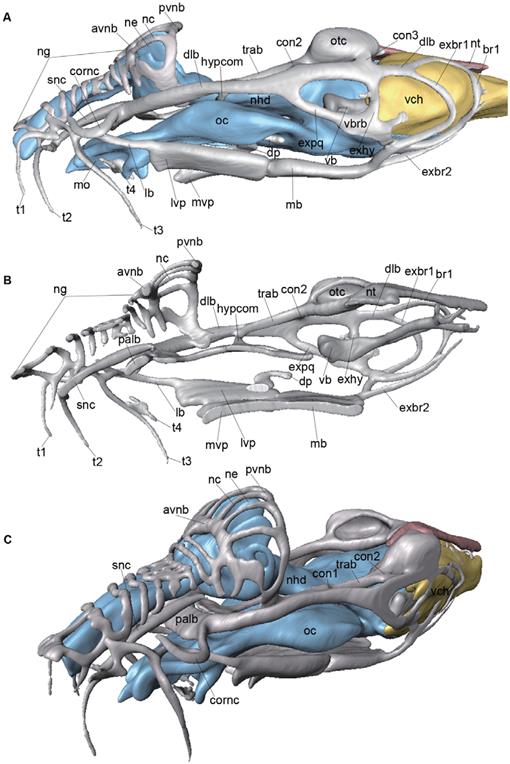
Fig. 8.
Chondrocranium of a prehatching-stage Eptatretus atami embryo. (B) Reconstruction of the chondrocranium. (A, C) Reconstruction of the chondrocranium with epithelial structures (ectodermal oronasohypophyseal cavity, light blue; pharyngeal endodermal lining, yellow). (A. B) lateral views. (C) Oblique anterior view. See the list in the text for abbreviations.
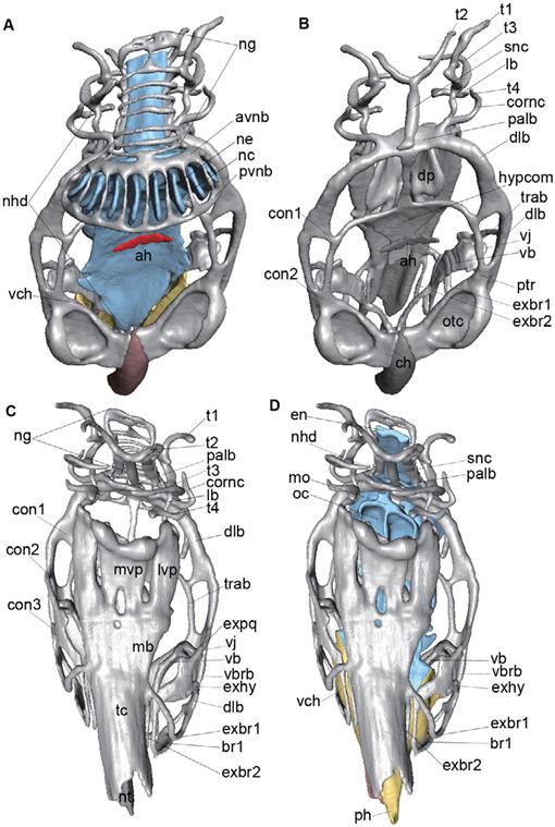
Fig. 9.
Chondrocranium of a prehatching-stage Eptatretus atami embryo. (A, C) Reconstruction of the chondrocranium. (B, D) Reconstruction of the chondrocranium with epithelial structures (ectodermal oronasohypophyseal cavity, light blue; pharyngeal endodermal lining, yellow). (A, B) ventral views (lingual plate is removed in (A)). (C , D) dorsal views. See the list in the text for abbreviations.
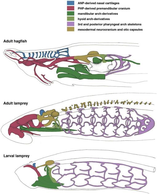
Fig. 10.
Homology of chondrocranial elements in cyclostomes. Hagfish and lamprey chondrocrania were compared on the basis of our results. Hagfish chondrocranium was redrawn from the work of Holmgren and Stensiö (1936), and those of the lamprey from the work of Marinelli and Strenger (1954) and Fontaine (1958).
Table 1.
Comparison of chondrocranial elements in cyclostomes.


