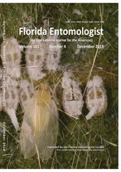
The sea grape flatid, Petrusa epilepsis (Kirkaldy) (Hemiptera: Flatidae) is a polyphagous, widespread planthopper in the Caribbean that infests common ornamental plants, as well as some of agricultural importance. In 2015, P. epilepsis was found in Florida for the first time, and was confirmed to be established in 2017 by the detection of eggs and nymphs on various ornamental plants located at the University of Florida's Fort Lauderdale Research and Education Center in Davie, Florida, USA. In this study, 9 new host plants are reported with 1 representing a new host family (Araliaceae). Additionally, the presence of adults, eggs, and nymphs of various sizes suggests that there are overlapping generations. Molecular analysis reveals no genetic variation of the COI gene. This study records establishment of another invasive species that has the potential to become a pest due to the large number of ornamental plants that are grown in South Florida that could serve as hosts of P. epilepsis elsewhere in Florida and the Caribbean.
The sea grape flatid, Petrusa epilepsis (Kirkaldy) (Hemiptera: Flatidae), is a common species of planthopper found throughout the Caribbean (Table 1) on a wide variety of host plants. This species is known from at least 37 host plants within 23 plant families (Table 2). On Guana Island (British Virgin Islands), P. epilepsis was found to be the most abundant planthopper sampled (Bartlett 2000). Petrusa epilepsis is not known to cause major economic losses. It is a common, minor pest of mostly ornamental plants, but also is found on crops of agricultural importance, such as coffee (Coffea arabica L.) (Rubiaceae) (Borkhataria et al. 2006) and mango (Mangifera indica L.) (Anacardiaceae) (S. H., unpublished). Feeding and subsequent honeydew accumulation from P. epilepsis has resulted in the growth of sooty mold on black mangroves (Avicennia germinans L.) Stearn (Acanthaceae) in Puerto Rico (Nieves–Rivera et al. 2012). Additionally, P. epilepsis was documented on the palms Cocos nucifera and Sabal causarum (both Arecaceae) in Puerto Rico (Segarra–Carmona et al. 2013). The common name of P. epilepsis is derived from its noted abundance on sea grape (Coccoloba uvifera L.) Jacq. (Polygonaceae) in its native range. Despite being widespread and abundant throughout the Caribbean, little is known about the biology of P. epilepsis, with the primary source of information on biology and life history being covered by Otero (2017).
The first evidence of P. epilepsis in Florida occurred in Jun 2015 when it was found in Hollywood, Florida, USA, by Florida Department of Agriculture and Consumer Services, Division of Plant Industry. Adults and immatures were found on Lantana involucrata (L.) (Verbenaceae) (Florida State Collection of Arthropods [FSCA] numbers E2015–2880, 3068), and adults and cast nymphal exoskeletons were found on Forestiera segregata (Jacq.) Krug & Urban (Oleaceae) (FSCA# E2015–3439). The insect was presumed to be established, since it was found on several plants of each species, but no further samples arrived at the Division of Plant Industry diagnostic bureau until fall 2017. No research has been conducted on P. epilepsis since its discovery in Florida, and information on host range, life history, genetic diversity, and distribution in Florida is lacking.
Table 1.
Distribution of Petrusa epilepsis (Hemiptera: Flatidae) in the Caribbean, excluding Florida.

In this study, evidence of the establishment of P. epilepsis in the US is presented, along with notes and observations on host plants, damage, and life history of a population collected at the University of Florida, Fort Lauderdale Research and Education Center, Fort Lauderdale, Florida, USA. Additionally, an evaluation of the genetic diversity of the local population is provided.
Materials and Methods
LOCALITY DATA, SAMPLING, AND IDENTIFICATION
This study was conducted at the Fort Lauderdale Research and Education Center, Davie, Broward County, Florida (26.050412°N, 80.141791°W), in the courtyard adjacent to the main administrative building. Initial samples collected were stored at −20 °C and then photographed using a Leica microscrope (Leica Microsystems, Buffalo Grove, Illinois, USA) and LASCore software (Leica Microsystems, Buffalo Grove, Illinois, USA). Male genitalia dissections were performed on representative males and identifications verified using Caldwell & Martorell (1951) and specimens from the Florida State Collection of Arthropods. The original find was confirmed by Dr. Charles Bartlett, University of Delaware (Newark, Delaware, USA). Following a modified protocol from Bahder et al. (2015), only the terminal end of the abdomen was used for both molecular and morphological analysis. After verification of the species identity, a survey was conducted at the Fort Lauderdale Research and Education Center on 24 Jan 2018 around the area where P. epilepsis was first observed. Additionally, all specimens of sea grape and mango on the Fort Lauderdale Research and Education Center property were visually inspected and sampled with sweep nets to determine the host range and local distribution.
DNA BARCODE FOR MOLECULAR CHARACTERIZATION
In order to develop a barcode and template for molecular characterization of P. epilepsis, the 5' region of COI was selected and amplified using the primers LCO1490 (forward) and HCO2198 (reverse). Primer sequences are presented in Table 3. Reactions were performed in 25 µl reaction consisting of 5X Clear GoTaq Flexi Buffer (Promega, Madison, Wisconsin, USA), 25 mM MgCl2, 200 µM dNTPs, 0.5 µM of each primer, 2% PVP–40, 1.5U GoTaq Flexi DNA polymerase (Promega, Madison, Wisconsin, USA), 2 µl of DNA template with the final volume prepared using Ultrapure (ThermoFisher Scientific, Waltham, Massachusetts, USA) sterile water. Thermal cycling conditions were as follows: initial denaturation at 95 °C for 2 min, followed by 35 cycles of denaturation at 95 °C for 30 sec, annealing at 50 °C for 30 sec, extension at 72 °C for 30 sec, followed by a final extension of 72 °C for 5 min. The amplicon was cloned using the TOPO TA Cloning Kit (ThermoFisher Scientific, Waltham, Massachusetts, USA) and the pCR®2.1 Vector (ThermoFisher Scientific, Waltham, Massachusetts, USA). After the cloned sequence was obtained, a total of 8 individuals were selected for molecular characterization and split based on color morph so that 4 individuals of each color were sequenced. PCR products were run on a 1.5% agarose gel stained with GelRed® (Biotium, Fremont, California, USA). All successful reactions were purified using ExoSAP–ITTM PCR Product Cleanup Reagent (ThermoFisher Scientific, Waltham, Massachusetts, USA) according to the manufacturer's instructions. Purified PCR product was quantified using a NanoDropLite spectrophotometer (ThermoFisher Scientific, Waltham, Massachusetts, USA) and sent for sequencing at Eurofins Scientific (Louisville, Kentucky, USA). Contiguous files were assembled using DNA Baser (Version 4.36) (Heracle BioSoft SRL, Pitesti, Romania), aligned using MEGA7 (Kumar et al. 2016).
Table 2.
Host range of Petrusa epilepsis (Hemiptera: Flatidae) in the Caribbean, excluding Florida.

Table 3.
Primers used to obtain COI barcoding information for Petrusa epilepsis (Hemiptera: Flatidae) in Florida.

Results
INITIAL OBSERVATIONS
The first adult P. epilepsis was observed at the Fort Lauderdale Research and Education Center on a glass door in the courtyard of the administrative building at the center on 28 Aug 2017. The individual was collected and photographed (Fig. 1). Other adults of both color morphs were found feeding on 2 different host plants in the courtyard: firebush (Hamelia patens Jacq.) (Rubiaceae) (Fig. 2), and blue porterweed (Stachytarpheta jamaicensis L. Vahl) (Verbenaceae) (Fig. 3). On this initial sample date, only adults were observed; however, both color morphs that have been reported for this species were found (Fig. 1). Following this, the population was monitored on a weekly basis from 1 Sep to 15 Dec 2017. The adult counts per wk for each plant are presented in Table 4. On 27 Sep 2017, egg masses were first observed on both H. patens (Fig. 4) and S. jamaicensis (Fig. 4). Nymphs were first observed on 1 Oct 2017, and were present through mid–Dec. Two types of damage were observed on the host plants. On infested H. patens, a general reddening of the leaves was apparent, while an adjacent H. patens that was not infested with P. epilepsis lacked these symptoms (Fig. 5). The infested S. jamaicensis, which had noticeably more egg masses and more nymphs, had sooty mold due to honeydew produced by nymphs (Fig. 5).
HOST SURVEY
The survey conducted in Jan 2018 revealed that 7 different plant species were fed upon by P. epilepsis (Table 5). Additional records from the Florida State Collection of Arthropods bring the list to 9 host plants for P. epilepsis in Florida. Five of the 7 hosts identified in the survey appeared to be reproductive hosts due to the presence of egg masses and nymphs (Table 5). No individuals were identified outside of the courtyard where they were first observed, and they appear to be very restricted in range at the center. All hosts recorded in this survey represent new host records for P. epilepsis and 1 new family record (Araliaceae).
MOLECULAR CHARACTERIZATION
The sequence obtained for the COI gene after cloning was a 709 base pair (bp) region from the 5' end (Accession No. MH041629). One record of P. epilepsis is present in GenBank (JN797344.1) for the COI gene; however the region available has an overlap of 96 base pairs with the product produced in this study, allowing for the sequences of other species that had a higher query coverage to come up after the BLAST search. The 96 bp overlap was 100% between the isolates. The closest match for the region sequenced in GenBank was from Metcalfa pruinosa (Say) (Hemiptera: Flatidae) (KX761467.1) with 87% identity and a query coverage of 68%. Based on the analysis performed, all 8 individuals had identical COI sequences. All isolates from this study have been deposited in GenBank (Accession Nos. MH041622–MH041628).
Discussion
This study provides the first evidence of the establishment of P. epilepsis in Florida. Additionally, it increases the known host range from 40 plant species in 23 families to 49 plant species in 24 families. This list represents confirmed feeding and reproductive hosts; however, the true range of reproductive hosts is unknown and needs further investigation. Future studies should focus on a survey of the urban environment to determine the distribution and host range of this species in South Florida. It is expected that in time the host range in Florida will increase. In addition, P. epilepsis is known to be a pest of ornamentals and some food crops in the Caribbean (Nieves–Rivera et al. 2012; Borkhataria et al. 2012), and may become a pest in Florida as well. After a better understanding of the distribution and host range of P. epilepsis in Florida is attained, applied studies for developing chemical and cultural practices to control populations will be undertaken. Due to the lack of information on the biology of this planthopper, it is unknown if natural enemies occur in Florida that could be used for biocontrol of this species. All damage recorded because of this species is limited to direct physical feeding, and the accumulation of honeydew and subsequent growth of sooty mold (Nieves–Rivera et al. 2012) for which the ornamental plant industry has a very low threshold. Until recently, no flatids were known to transmit plant pathogens (Wilson & O'Brien 1987); however, Donati et al. (2017) have shown that M. pruinosa Say can transmit Pseudomonas syringae pv. actinidiae van Hall (Pseudomonadaceae) to healthy kiwi (Actinidia spp.) plants. It is unknown if P. epilepsis is capable of transmitting plant pathogens, but due to its phloem–feeding behavior, it could play a role in transmission of known or unknown plant pathogens. Because it is polyphagous and newly established, this is an aspect of its biology that should be investigated further.
Sources for distribution and early host records were obtained from Metcalf (1957). Records from South and Central America are poorly supported. The only record from Brazil is from Stål (1869), with no details given. The record from Brazil in Melichar (1902, 1923) references Stål. Similarly, “Insulae Americanae Meridionalis,” South American islands, in Stål (1869) could refer to Caribbean islands, as does Melichar's (1923) reference to Central America, where St. Croix, St. Thomas, and Barthélemy are specified. Metcalf (1957) says that Melichar (1923) records P. epilepsis in Colombia, but we could not find any reference to Colombia in Melichar (1923).
Table 4.
Population data of Petrusa epilepsis (Hemiptera: Flatidae) collected from the Hamelia patens and Stachytarpheta jamaicensis in 2017.

Whereas only 8 specimens were analyzed at the molecular level, the development of a barcode for this species provides information for future molecular studies to determine the genetic variability within the Florida population. A larger number of individuals from the Fort Lauderdale Research and Education Center needs to be analyzed, as well as individuals from other Florida localities, assuming it is established elsewhere. Additionally, further analysis of specimens from the Caribbean will provide information on genetic variation within its native range and could be used to determine the origin of this species. Since so few individuals were analyzed, it is unknown if there is a genetic basis for the color morphs or if the variants are due to environmental conditions. Regardless, the fact that both color morphs have identical sequences supports the morphological work done by Fennah (1941) indicating that they are the same species.
Fig. 4.
Egg masses of Petrusa epilepsis on Stachytarpheta jamaicensis (blue porterweed) (A) and Hamelia patens (firebush) (B, C).

Fig. 5.
Feeding damage of Petrusa epilepsis on Stachytarpheta jamaicensis (blue porterweed) (A) and Hamelia patens (firebush) (B).

Table 5.
Host plants of Petrusa epilepsis (Hemiptera: Flatidae) recorded from first detection1 and from the survey conducted in Jan 2018 at the University of Florida, Fort Lauderdale Research and Education Center, Davie, Florida2.

This study provides evidence of the establishment of a new invasive species in Florida and a baseline for studying its ecology and invasion biology. It also highlights that non–native species are continually being detected in Florida, emphasizing the importance of improved identification protocols.
Acknowledgments
We thank Charles Bartlett, Department of Entomology and Wildlife Ecology, University of Delaware, for help identifying the first specimens and critical review of the manuscript. We also thank Sarah Kern and Susan Thor, both University of Florida, Fort Lauderdale Research and Education Center, Fort Lauderdale, Florida, USA, for providing valuable information regarding host plants. We thank Jeffrey Eby, Florida Department of Agriculture and Consumer Services, Division of Plant Industry, Gainesville, for library assistance and Patti Anderson, Florida Department of Agriculture and Consumer Services, Division of Plant Industry, for botanical review. We thank Cristina Urbina and Kevin Williams, Florida Department of Agriculture and Consumer Services, Division of Plant Industry inspectors, for assistance in conducting the host survey. Finally, we thank William Kern, University of Florida, Fort Lauderdale Research and Education Center, Fort Lauderdale, Florida, USA, and Stephen Wilson, Department of Biology and Agriculture, University of Central Missouri, Warrensburg, Missouri, USA, for initial review of the manuscript.


